. 2015 Feb 12;6(2):e1636.
doi: 10.1038/cddis.2015.16.

Differential roles of RIPK1 and RIPK3 in TNF-induced necroptosis and chemotherapeutic agent-induced cell death

Affiliations

Differential roles of RIPK1 and RIPK3 in TNF-induced necroptosis and chemotherapeutic agent-induced cell death

K Moriwaki et al. Cell Death Dis. .

Abstract

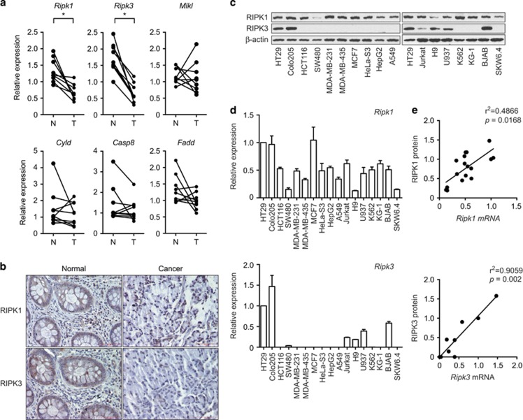
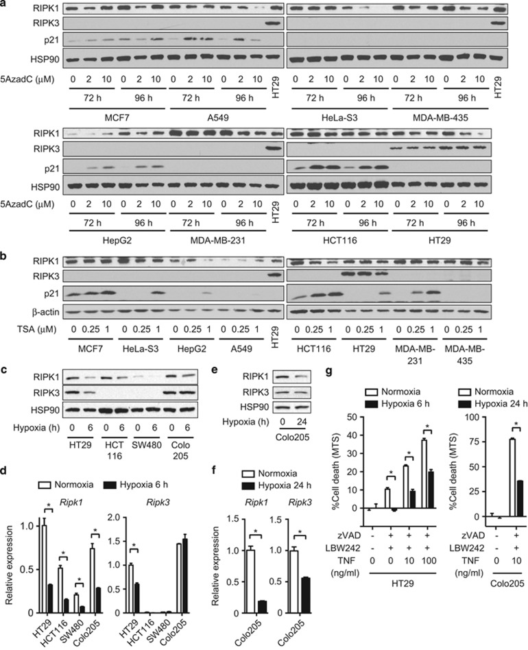
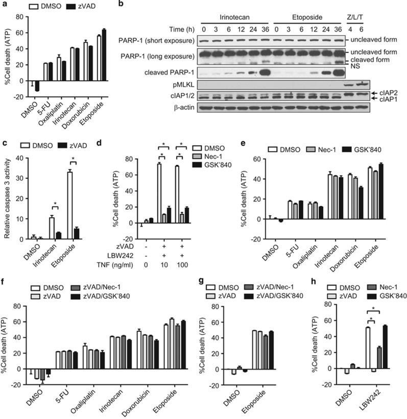
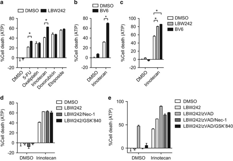
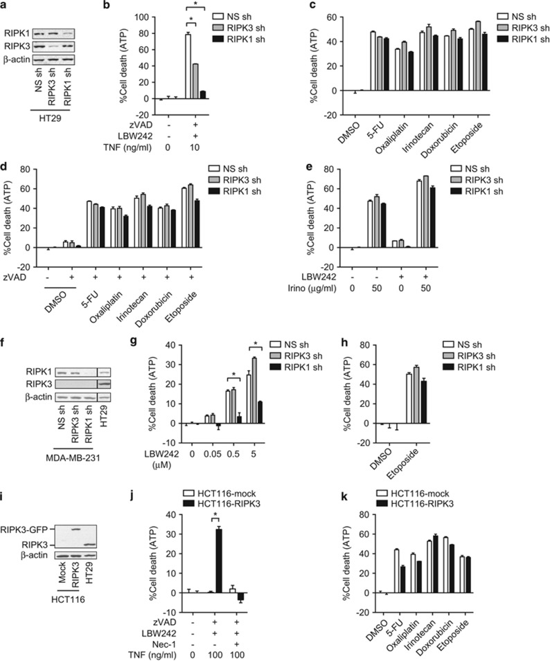
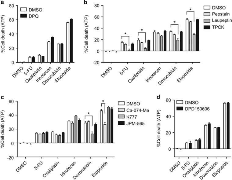
Apoptosis is a key mechanism for metazoans to eliminate unwanted cells. Resistance to apoptosis is a hallmark of many cancer cells and a major roadblock to traditional chemotherapy. Recent evidence indicates that inhibition of caspase-dependent apoptosis sensitizes many cancer cells to a form of non-apoptotic cell death termed necroptosis. This has led to widespread interest in exploring necroptosis as an alternative strategy for anti-cancer therapy. Here we show that in human colon cancer tissues, the expression of the essential necroptosis adaptors receptor interacting protein kinase (RIPK)1 and RIPK3 is significantly decreased compared with adjacent normal colon tissues. The expression of RIPK1 and RIPK3 was suppressed by hypoxia, but not by epigenetic DNA modification. To explore the role of necroptosis in chemotherapy-induced cell death, we used inhibitors of RIPK1 or RIPK3 kinase activity, and modulated their expression in colon cancer cell lines using short hairpin RNAs. We found that RIPK1 and RIPK3 were largely dispensable for classical chemotherapy-induced cell death. Caspase inhibitor and/or second mitochondria-derived activator of caspase mimetic, which sensitize cells to RIPK1- and RIPK3-dependent necroptosis downstream of tumor necrosis factor receptor-like death receptors, also did not alter the response of cancer cells to chemotherapeutic agents. In contrast to the RIPKs, we found that cathepsins are partially responsible for doxorubicin or etoposide-induced cell death. Taken together, these results indicate that traditional chemotherapeutic agents are not efficient inducers of necroptosis and that more potent pathway-specific drugs are required to fully harness the power of necroptosis in anti-cancer therapy.


Conflict of interest statement

JB and PJ are employees of GlaxoSmithKline. All other authors declare no potential conflict of interest.

Figures

Figure 1
Figure 1
The expression of Ripk1 and Ripk3 is decreased in human colon cancer. (a) Total RNA from human colon cancer tissues (T) and adjacent normal colon tissues (N) were analyzed by real-time PCR for the expression of Ripk1, Ripk3, Mlkl, Cyld, Casp8, and Fadd. P-values were calculated by Wilcoxon matched-pairs signed-rank test. *P<0.05. (b) Pictures of normal and cancer tissues stained for RIPK1 and RIPK3 are shown. Bars: 75 μM. (c) Whole-cell extracts and (d) RNA from various cancer cell lines were analyzed by western blotting and real-time PCR, respectively, for the expression of RIPK1 and RIPK3. For comparison between different cell lines, we define the expression in HT29 cells as 1. (e) Comparison of mRNA and protein level of RIPK1 (upper panel) and RIPK3 (lower panel). The intensity of RIPK1 and RIPK3 protein expression in c was quantified using ImageJ. The protein expression level in HT29 cells was defined as 1. P-value and r2 were determined by Spearman correlation coefficients. Error bars indicate S.E.M. (n=3)
Figure 2
Figure 2
RIPK1 and RIPK3 expression is regulated by hypoxia, but not by DNA methylation or histone deacetylation. (a) The cancer cell lines were treated with 5AzadC as indicated. (b) The cells were treated with TSA for 24 h. RIPK1, RIPK3, and p21 expression was determined by western blotting. (c–f) Cells were exposed to 1% O2 hypoxic condition for (c and d) 6 or (e and f) 24 h. (c and e) Whole-cell extracts and (d and f) RNA were prepared for western blotting and Q-PCR, respectively. (g) Necroptosis was induced by pretreatment with 20 μM zVAD and 10 μM LBW242 for 1 h, followed by treatment with TNF overnight. For hypoxia, HT29 cells and Colo205 cells were cultured in 1% O2 hypoxic condition for 6 and 24 h before and during necroptosis induction, respectively. Cell death was determined using CellTiter96 Aqueous Non-Radioactive Cell Proliferation Assay. Error bars indicate S.E.M. (n=3). *P<0.05
Figure 3
Figure 3
Chemotherapeutic agents induce caspase-independent non-necroptotic cell death. (a) HT29 cells were pretreated with zVAD before treatment with various chemotherapeutic agents. (b) Whole-cell extracts were prepared from HT29 cells treated with irinotecan or etoposide, and analyzed by western blotting. Necroptosis was induced by zVAD, LBW242, and TNF (Z/L/T). (c) Caspase 3 activity was quantified as described in Materials and Methods, in HT29 cells treated with irinotecan or etoposide for 36 h in the absence or the presence of zVAD. (d and e) Cell death was induced by (d) zVAD, LBW242, and TNF, or (e) the indicated chemotherapeutic agents in the absence or presence of Nec-1 or GSK'840 in HT29 cells. (f) HT29 and (g) MDA-MB-231 cells were pretreated with zVAD in combination with Nec-1 or GSK'840 before treatment with the chemotherapeutic agents. (h) MDA-MB-231 cells were pretreated with zVAD in combination with Nec-1 or GSK'840, and subsequently treated with 2.5 μM LBW242 for 48 h. Cell death was determined using CellTiter-Glo Luminescent Cell Viability Assay in a and d–h. Error bars indicate S.E.M. (n=3). *P<0.05
Figure 4
Figure 4
Smac mimetics enhance cancer cell death in a chemotherapeutic agent-specific manner. (a and b) HT29 cells or (c) Colo205 cells were pretreated with (a and c) 2.5 μM LBW242 or (b and c) 0.2 μM BV6 for 1 h and subsequently treated with chemotherapeutic agents. (d) HT29 cells pretreated with 2.5 μM LBW242 in the presence of Nec-1 or GSK'840 were treated with chemotherapeutic agents. (e) Cells were treated as in d, except that zVAD was added where indicated. Cell death was determined using CellTiter-Glo Luminescent Cell Viability Assay. Error bars indicate S.E.M. (n=3). *P<0.05
Figure 5
Figure 5
RIPK1 and RIPK3 are dispensable for chemotherapeutic agent-induced cell death. (a) Knockdown of RIPK1 or RIPK3 by shRNA in HT29 cells was confirmed by western blotting. (b) Necroptosis was induced by TNF, zVAD, and LBW242. (c and d) RIPK1 or RIPK3-knockdown HT29 cells were treated with the chemotherapeutic agents in the (c) absence or (d) presence of zVAD. (e) RIPK1 or RIPK3-knockdown HT29 cells were pretreated with 2.5 μM LBW242 for 1 h before irinotecan treatment. (f) RIPK1 and RIPK3 expression in MDA-MB-231 cells stably transfected with shRNA against RIPK1 or RIPK3. The lanes were run on the same gel but were non-contiguous. (g and h) RIPK1 or RIPK3-knockdown MDA-MB-231 cells were treated with (g) LBW242 or (h) etoposide for 48 h. (i) Overexpression of RIPK3-GFP in HCT116 cells was confirmed by western blotting. (j) Necroptosis was induced by TNF, zVAD-fmk, and LBW242 in RIPK3-GFP-expressing HCT116 cells. (k) RIPK3-GFP-expressing HCT116 cells were treated with the indicated chemotherapeutic agents. Cell death was determined using CellTiter-Glo Luminescent Cell Viability Assay. Error bars indicate S.E.M. (n=3). *P<0.05
Figure 6
Figure 6
Doxorubicin and etoposide induced cathepsin-dependent cell death. HT29 cells were pretreated with (a) 10 μM DPQ, (b) 1 μM pepstatin, 10 μM leupeptin, or 10 μM TPCK, (c) 10 μM Ca-074-Me, 10 μM K777, or 10 μM JPM-565, or (d) 10 μM PD150606 for 1 h before treatment with the chemotherapeutic agents. Cell death was determined using CellTiter-Glo Luminescent Cell Viability Assay. Error bars indicate S.E.M. (n=3). *P<0.05

Similar articles

Cited by

References

    1. 1Hanahan D, Weinberg RA. Hallmarks of cancer: the next generation. Cell 2011; 144: 646–674. - PubMed
    1. 2Alison MR, Lin WR, Lim SM, Nicholson LJ. Cancer stem cells: in the line of fire. Cancer Treat Rev 2012; 38: 589–598. - PubMed
    1. 3Kroemer G, Galluzzi L, Kepp O, Zitvogel L. Immunogenic cell death in cancer therapy. Annu Rev Immunol 2013; 31: 51–72. - PubMed
    1. 4Moriwaki K, Chan FK. RIP3: a molecular switch for necrosis and inflammation. Genes Dev 2013; 27: 1640–1649. - PMC - PubMed
    1. 5Li J, McQuade T, Siemer AB, Napetschnig J, Moriwaki K, Hsiao YS et al. The RIP1/RIP3 necrosome forms a functional amyloid signaling complex required for programmed necrosis. Cell 2012; 150: 339–350. - PMC - PubMed

Publication types

MeSH terms