

Spectroscopic and mutagenesis studies of human PGRMC1
- PMID: 25675345
- PMCID: PMC4533898
- DOI: 10.1021/bi501177e
Spectroscopic and mutagenesis studies of human PGRMC1
Abstract
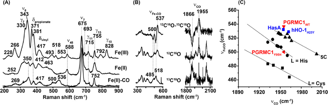
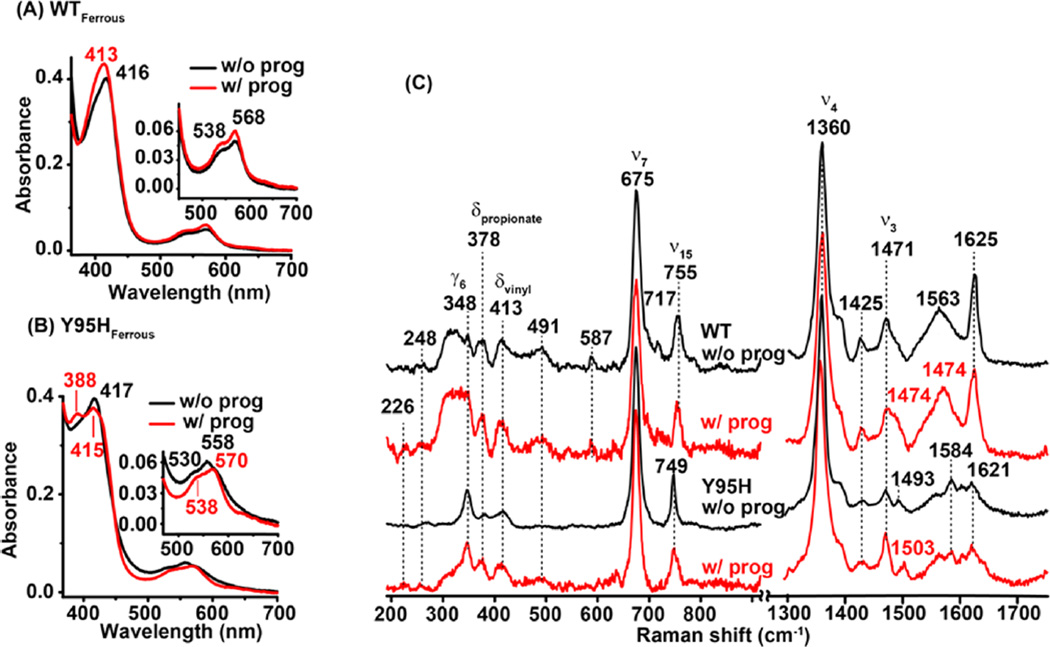
Progesterone receptor membrane component 1 (PGRMC1) is a 25 kDa protein with an N-terminal transmembrane domain and a putative C-terminal cytochrome b5 domain. Heme-binding activity of PGRMC1 has been shown in various homologues of PGRMC1. Although the general definition of PGRMC1 is as a progesterone receptor, progesterone-binding activity has not been directly demonstrated in any of the purified PGRMC1 proteins fully loaded with heme. Here, we show that the human homologue of PGRMC1 (hPGRMC1) binds heme in a five-coordinate (5C) high-spin (HS) configuration, with an axial tyrosinate ligand, likely Y95. The negatively charged tyrosinate ligand leads to a relatively low redox potential of approximately -331 mV. The Y95C or Y95F mutation dramatically reduces the ability of the protein to bind heme, supporting the assignment of the axial heme ligand to Y95. On the other hand, the Y95H mutation retains ∼90% of the heme-binding activity. The heme in Y95H is also 5CHS, but it has a hydroxide axial ligand, conceivably stabilized by the engineered-in H95 via an H-bond; CO binding to the distal ligand-binding site leads to an exchange of the axial ligand to a histidine, possibly H95. We show that progesterone binds to hPGRMC1 and introduces spectral changes that manifest conformational changes to the heme. Our data offer the first direct evidence supporting progesterone-binding activity of PGRMC1.
Figures


Similar articles
-
Progesterone receptor membrane component 1 (PGRMC1) binds and stabilizes cytochromes P450 through a heme-independent mechanism.J Biol Chem. 2021 Nov;297(5):101316. doi: 10.1016/j.jbc.2021.101316. Epub 2021 Oct 20. J Biol Chem. 2021. PMID: 34678314 Free PMC article.
-
Progesterone receptor membrane component-1 (PGRMC1) is the mediator of progesterone's antiapoptotic action in spontaneously immortalized granulosa cells as revealed by PGRMC1 small interfering ribonucleic acid treatment and functional analysis of PGRMC1 mutations.Endocrinology. 2008 Feb;149(2):534-43. doi: 10.1210/en.2007-1050. Epub 2007 Nov 8. Endocrinology. 2008. PMID: 17991724 Free PMC article.
-
The emerging role of progesterone receptor membrane component 1 (PGRMC1) in cancer biology.Biochim Biophys Acta. 2016 Dec;1866(2):339-349. doi: 10.1016/j.bbcan.2016.07.004. Epub 2016 Jul 22. Biochim Biophys Acta. 2016. PMID: 27452206 Review.
-
Cystathionine β-synthase and PGRMC1 as CO sensors.Free Radic Biol Med. 2016 Oct;99:333-344. doi: 10.1016/j.freeradbiomed.2016.08.025. Epub 2016 Aug 24. Free Radic Biol Med. 2016. PMID: 27565814 Review.
-
Defining Requirements for Heme Binding in PGRMC1 and Identifying Key Elements that Influence Protein Dimerization.Biochemistry. 2024 Apr 2;63(7):926-938. doi: 10.1021/acs.biochem.3c00718. Epub 2024 Mar 15. Biochemistry. 2024. PMID: 38489495
Cited by
-
PGRMC1: An enigmatic heme-binding protein.Pharmacol Ther. 2023 Jan;241:108326. doi: 10.1016/j.pharmthera.2022.108326. Epub 2022 Dec 1. Pharmacol Ther. 2023. PMID: 36463977 Free PMC article. Review.
-
Progesterone Receptor Membrane Component (PGRMC)1 and PGRMC2 and Their Roles in Ovarian and Endometrial Cancer.Cancers (Basel). 2021 Nov 26;13(23):5953. doi: 10.3390/cancers13235953. Cancers (Basel). 2021. PMID: 34885064 Free PMC article. Review.
-
Progesterone receptor membrane component 1 promotes survival of human breast cancer cells and the growth of xenograft tumors.Cancer Biol Ther. 2016;17(3):262-71. doi: 10.1080/15384047.2016.1139240. Epub 2016 Jan 19. Cancer Biol Ther. 2016. PMID: 26785864 Free PMC article.
-
Haem-dependent dimerization of PGRMC1/Sigma-2 receptor facilitates cancer proliferation and chemoresistance.Nat Commun. 2016 Mar 18;7:11030. doi: 10.1038/ncomms11030. Nat Commun. 2016. PMID: 26988023 Free PMC article.
-
An Analysis of the Multifaceted Roles of Heme in the Pathogenesis of Cancer and Related Diseases.Cancers (Basel). 2021 Aug 17;13(16):4142. doi: 10.3390/cancers13164142. Cancers (Basel). 2021. PMID: 34439295 Free PMC article. Review.
References
-
- Min L, Strushkevich NV, Harnastai IN, Iwamoto H, Gilep AA, Takemori H, Usanov SA, Nonaka Y, Hori H, Vinson GP. Molecular identification of adrenal inner zone antigen as a heme-binding protein. FEBS J. 2005;272:5832–5843. - PubMed
-
- Raza FS, Takemori H, Tojo H, Okamoto M, Vinson GP. Identification of the rat adrenal zona fasciculata/reticularis specific protein, inner zone antigen (IZAg), as the putative membrane progesterone receptor. Eur. J. Biochem. 2001;268:2141–2147. - PubMed
-
- Min L, Takemori H, Nonaka Y, Katoh Y, Doi J, Horike N, Osamu H, Raza FS, Vinson GP, Okamoto M. Characterization of the adrenal-specific antigen IZA (inner zone antigen) and its role in the steroidogenesis. Mol. Cell. Endocrinol. 2004;215:143–148. - PubMed
-
- Lösel RM, Besong D, Peluso JJ, Wehling M. Progesterone receptor membrane component 1–many tasks for a versatile protein. Steroids. 2008;73:929–934. - PubMed
-
- Selmin O, Lucier GW, Clark GC, Tritscher AM, Heuvel JPV, Gastel JA, Walker NJ, Thomas R, Bell DA. Isolation and characterization of a novel gene induced by 2, 3, 7, 8-tetrachlorodibenzo-p-dioxin in rat liver. Carcinogenesis. 1996;17:2609–2615. - PubMed
Publication types
MeSH terms
Substances
Grants and funding
LinkOut - more resources
Full Text Sources
Other Literature Sources
Molecular Biology Databases
Research Materials
