. 2015 Feb 24;112(8):2425-30.
doi: 10.1073/pnas.1411514112. Epub 2015 Feb 9.

In-cell SHAPE reveals that free 30S ribosome subunits are in the inactive state

Affiliations

In-cell SHAPE reveals that free 30S ribosome subunits are in the inactive state

Jennifer L McGinnis et al. Proc Natl Acad Sci U S A. .

Abstract

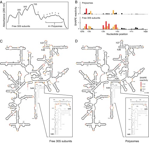
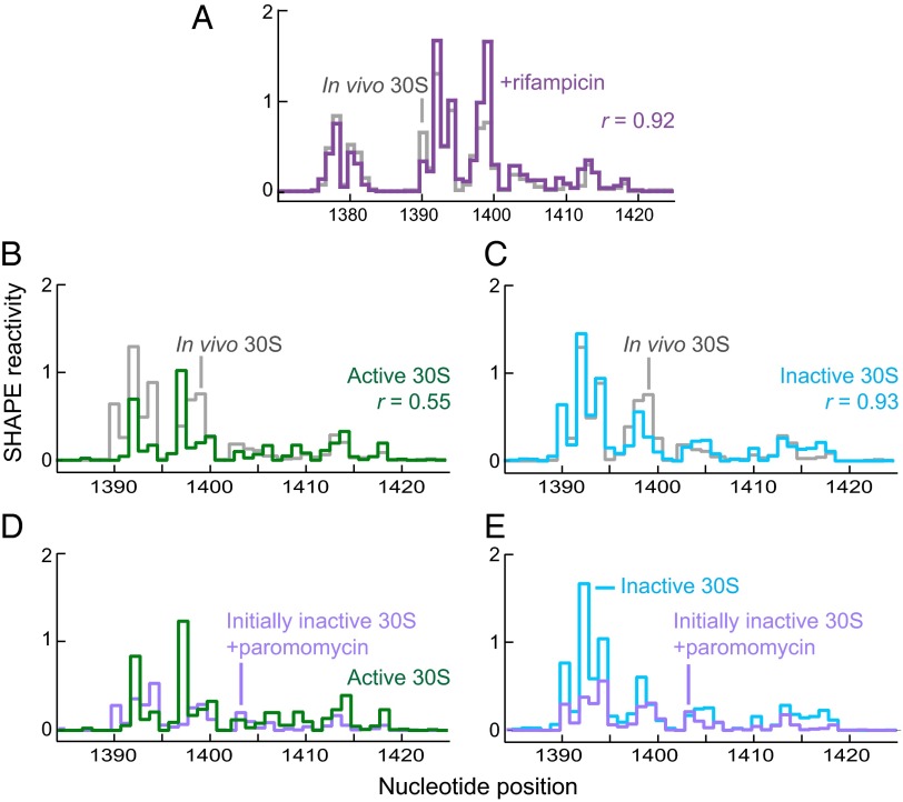
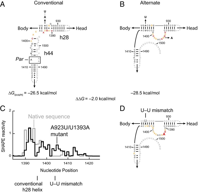
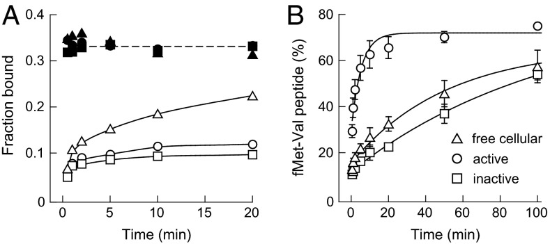
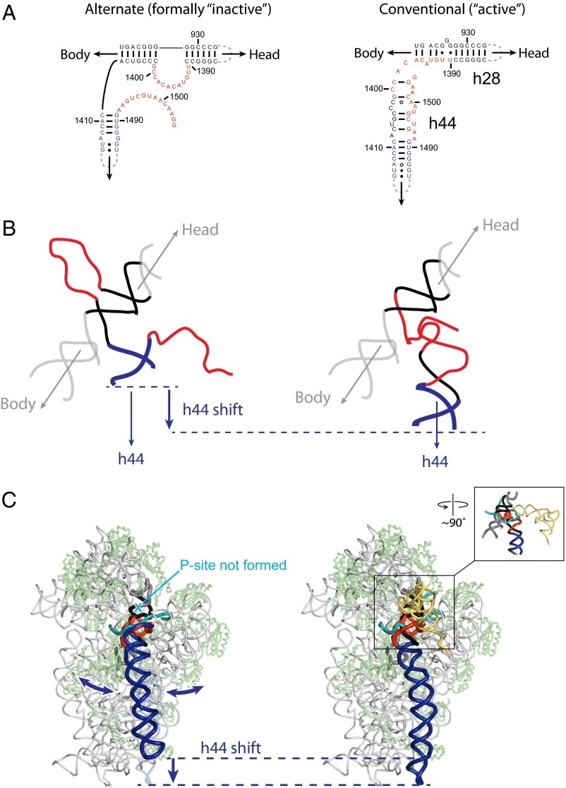
It was shown decades ago that purified 30S ribosome subunits readily interconvert between "active" and "inactive" conformations in a switch that involves changes in the functionally important neck and decoding regions. However, the physiological significance of this conformational change had remained unknown. In exponentially growing Escherichia coli cells, RNA SHAPE probing revealed that 16S rRNA largely adopts the inactive conformation in stably assembled, mature 30S subunits and the active conformation in translating (70S) ribosomes. Inactive 30S subunits bind mRNA as efficiently as active subunits but initiate translation more slowly. Mutations that inhibited interconversion between states compromised translation in vivo. Binding by the small antibiotic paromomycin induced the inactive-to-active conversion, consistent with a low-energy barrier between the two states. Despite the small energetic barrier between states, but consistent with slow translation initiation and a functional role in vivo, interconversion involved large-scale changes in structure in the neck region that likely propagate across the 30S body via helix 44. These findings suggest the inactive state is a biologically relevant alternate conformation that regulates ribosome function as a conformational switch.

Keywords: 16S rRNA; SHAPE; conformational change; in vivo; ribosome.


Conflict of interest statement

The authors declare no conflict of interest.

Figures

Fig. 1.
Fig. 1.
In vivo SHAPE analysis of ribosome complexes. (A) Ribosome complexes from E. coli cells, probed with 1M7, partitioned on sucrose density gradients. Peaks corresponding to 30S and 50S ribosomal subunits, 70S ribosomes, and polysomes containing four to eight ribosomes are indicated; the top of the gradient is on the left. (B) SHAPE reactivity profiles for 16S rRNA from polysomes (Top) and free 30S subunits (Bottom). (C and D) SHAPE reactivities for 16S rRNA isolated from (C) free 30S subunits and (D) polysomes superimposed on the conventional secondary structure. Nucleotides are shown as circles, colored by SHAPE reactivity (see scale). As shown in the Insets, reactivities differ in the neck region, and reactivities for the 16S rRNA in free 30S subunits are inconsistent with the conventional structure.
Fig. 2.
Fig. 2.
16S rRNA structure in 30S subunits in vivo resembles the in vitro inactive state, and paromomycin induces the switch from inactive to active conformation. (A) Histograms comparing in-cell SHAPE reactivities for 16S rRNA from free 30S subunits isolated from cells during log-phase growth (gray) with 16S rRNA reactivities from free 30S subunits isolated from cells treated with rifampicin (purple). (B and C) Histograms comparing in-cell SHAPE reactivities for 16S rRNA isolated from 30S subunits (gray) with reactivities for 16S rRNA from isolated 30S subunits treated in vitro under conditions that yield (B) active (green) and (C) inactive (cyan) 30S subunits. (D and E) Histograms comparing SHAPE reactivities for 16S rRNA from inactive 30S subunits treated in vitro with paromomycin (purple) with those from subunits obtained in vitro under conditions that yield (D) active (green) and (E) inactive (cyan) 30S subunits.
Fig. 3.
Fig. 3.
Alternate secondary structure for 16S rRNA in free 30S subunits in vivo. (A) Conventional base-pairing model and (B) SHAPE-supported alternate model (3) of 16S rRNA. SHAPE reactivity profile for 16S rRNA from free 30S subunits isolated after in vivo SHAPE probing are superimposed on each structure model. (C) Histograms comparing 16S rRNA SHAPE reactivities for the native sequence (gray) and A923U/U1393A mutant (black) from free 30S subunits isolated after in vivo modification. Mutant-specific structural landmarks are shown. (D) SHAPE reactivities for the mutant 16S rRNA superimposed on the alternate base-pairing model. The two mutated nucleotides are shown in a larger font. The experiments shown in A and B versus C and D were performed on wild-type and ∆7 prrn E. coli cells, respectively; a full set of comparisons showing SHAPE reactivities superimposed on both conventional and alternate secondary structure models are shown in Figs. S2 and S5. Nucleotides are colored by SHAPE reactivity using the red, yellow, and black scale shown in Fig. 1.
Fig. 4.
Fig. 4.
mRNA binding and translation initiation activities of 30S subunits. (A) mRNA binding, assayed by nitrocellulose filter binding. poly(U) and m292 mRNAs are shown in filled and open symbols, respectively. (B) Translation initiation measured by dipeptide formation. Data were fit to a single-exponential function; for active, inactive, and free cellular 30S subunits, kapp was 0.38 ± 0.10, 0.0093 ± 0.0012, and 0.020 ± 0.005 min–1, respectively. Error bars show the SEM from three or more independent experiments.
Fig. 5.
Fig. 5.
Structural and mechanistic consequences for formation of the alternate secondary structure in the 16S rRNA neck helices. (A) Secondary structures for alternate and conventional models of the 16S rRNA neck helices. (B) 3D models for the alternate (Left) and conventional (Right) structures. The alternate and conventional models were based on discrete molecular dynamics and crystallographic structures (26), respectively. (C) Illustration of the position for h44 in the context of the 30S subunit for the alternate and conventional conformations of the 16S rRNA. In the diagram of the alternate state, double-headed arrows emphasize likely conformational dynamics of h44. Inset illustrates area highlighted in B plus tRNA (yellow) and mRNA (cyan).

References

    1. Zamir A, Miskin R, Elson D. Interconversions between inactive and active forms of ribosomal subunits. FEBS Lett. 1969;3(1):85–88. - PubMed
    1. Moazed D, Van Stolk BJ, Douthwaite S, Noller HF. Interconversion of active and inactive 30 S ribosomal subunits is accompanied by a conformational change in the decoding region of 16 S rRNA. J Mol Biol. 1986;191(3):483–493. - PubMed
    1. Lavender CA, et al. Model-free RNA sequence and structure alignment informed by SHAPE probing reveals a conserved alternate secondary structure for 16S rRNA. PLoS Comp Biol. 2015 in press. - PMC - PubMed
    1. Swiatkowska A, et al. Kinetic analysis of pre-ribosome structure in vivo. RNA. 2012;18(12):2187–2200. - PMC - PubMed
    1. McGinnis JL, Weeks KM. Ribosome RNA assembly intermediates visualized in living cells. Biochemistry. 2014;53(19):3237–3247. - PMC - PubMed

Publication types

MeSH terms