. 2015 Feb 24;112(8):2419-24.
doi: 10.1073/pnas.1419582112. Epub 2015 Feb 9.

Visualization of a radical B12 enzyme with its G-protein chaperone

Affiliations

Visualization of a radical B12 enzyme with its G-protein chaperone

Marco Jost et al. Proc Natl Acad Sci U S A. .

Abstract

G-protein metallochaperones ensure fidelity during cofactor assembly for a variety of metalloproteins, including adenosylcobalamin (AdoCbl)-dependent methylmalonyl-CoA mutase and hydrogenase, and thus have both medical and biofuel development applications. Here, we present crystal structures of IcmF, a natural fusion protein of AdoCbl-dependent isobutyryl-CoA mutase and its corresponding G-protein chaperone, which reveal the molecular architecture of a G-protein metallochaperone in complex with its target protein. These structures show that conserved G-protein elements become ordered upon target protein association, creating the molecular pathways that both sense and report on the cofactor loading state. Structures determined of both apo- and holo-forms of IcmF depict both open and closed enzyme states, in which the cofactor-binding domain is alternatively positioned for cofactor loading and for catalysis. Notably, the G protein moves as a unit with the cofactor-binding domain, providing a visualization of how a chaperone assists in the sequestering of a precious cofactor inside an enzyme active site.

Keywords: G protein; crystallography; metallochaperone; metallocofactor delivery; vitamin B12.


Conflict of interest statement

The authors declare no conflict of interest.

Figures

Fig. 1.
Fig. 1.
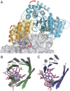
Crystal structure of IcmF and comparison with MCM/MMAA. (A) Dimeric IcmF in ribbon depiction, colored by domain with one chain lighter than the other: yellow (Cbl-domains), cyan (G-domains), green (substrate-binding domains), pink/red (structured linker regions). AdoCbl shown in ball-and-stick with Cbl carbon in pink, 5′-dAdo carbon in cyan, cobalt in purple; GDP•Mg2+ in ball-and-stick with carbon in yellow, Mg2+ in orange. Boxed region shown in more detail in Fig. 2A. (B and C) IcmF protomer in a different orientation, with (B) mutase domains shown slightly separated from (C) the G-domain to emphasize shape complementarity. The second IcmF protomer and linker regions are not shown for clarity. (D) Human MCM protomer [PDB ID code 2XIQ (33)] and (E) dimeric MMAA [PDB ID code 2WWW (33)], shown in the same orientation as the corresponding IcmF domains in (B) and (C). MCM is colored in blue (substrate-binding domain) and red (Cbl-binding domain). The second MCM protomer is not shown for clarity. The MMAA protomers are shown in cyan and gray. MCM-bound Cbl and MMAA-bound GDP are shown as in A. (F) Close-up of IcmF domain interactions, with G- and Cbl-domains shown as in A and the substrate-binding domain in surface representation. (G) Structure-based docking model of the human MCM:MMAA complex, generated by superimposition of human MCM [PDB ID code 2XIQ (33)] and MMAA [PDB ID code 2WWW (33)] structures onto IcmF and oriented as in F. MCM shown with substrate-binding domain in surface representation and Cbl-domain in red, MMAA shown in cyan, Cbl and GDP shown as in A. Switch I of MMAA is disordered, locations of the chain breaks are indicated by spheres and connected by dashed lines.
Fig. 2.
Fig. 2.
Molecular interactions within and between IcmF domains. (A) IcmF GTPase active site with signature G-protein elements highlighted: Mg2+-binding P-loop or Walker A motif (purple); switch I (dark blue), which is conformationally responsive to the GTPase cycle; switch II or Walker B motif (pink), which also undergoes conformational changes; and base specificity loop (orange) with the sequence NKxD that confers specificity to guanosine nucleotides. GDP•Mg2+ shown with carbon in yellow, Mg2+ in orange. Hydrogen bonds and coordination bonds are indicated by dashed black lines. Water molecules may additionally coordinate the Mg2+, but are not visible in this structure because of its modest resolution. (B) Close-up view of the interactions between selected parts of the G-domain (cyan) and mutase Cbl- (yellow, orange) and substrate-binding domains (green), oriented as Fig. 1F. AdoCbl shown with Cbl carbon in pink, 5′-dAdo carbon in cyan, cobalt in purple. Switch I shown in dark blue. Interactions between G-domain and mutase domains are shown as black dashed lines, with residues shown as sticks with carbons colored by domains. Pathogenic mutations in MCM:MMAA, which have been mapped onto IcmF, are highlighted in pink (R28, G54, L60, G257). (C and D) Close-up view of mapped pathogenic mutations; see Fig. S4B for location in overall structure. IcmF, colored as in B, is superimposed with MMAA in gray. Highlighted pathogenic mutations are (C) Arg28 (MCM Arg616) as well as (D) Gly54 (MCM Gly642) and Gly257 (MMAA Gly188), shown in pink. In D, the corresponding region in MMAA is disordered and not shown.
Fig. 3.
Fig. 3.
Conformational movements in IcmF. (A) Differential positioning of the Cbl-domain and G-domain of holo-IcmF•GDP in chain A (cyan/orange) compared with chain B (light blue/yellow) when superimposed by their substrate-binding domains (gray surfaces). Cbl is shown in pink (chain A) and black (chain B), and GDP in yellow (chain A) and green (chain B). Mg2+ is shown as an orange sphere. 5′-dAdo not shown for clarity. (B) Superposition of the TIM barrel β-strands of holo-IcmF•GDP chains A (dark green) and B (gray). Cbl colored as in A and distances between corresponding Cα-atoms in Å. (C) Superposition of the TIM barrel β-strands of substrate-bound (dark blue) and substrate-free (gray) MCM [PDB ID codes 4REQ (35) and 2REQ (27), respectively]. Cbl is shown in pink (substrate-bound) and black (substrate-free).
Fig. 4.
Fig. 4.
Schematic of proposed molecular model of Cbl delivery. Only one monomer of the dimer is depicted. See main text for details.

Similar articles

Cited by

References

    1. Tottey S, Harvie DR, Robinson NJ. Understanding how cells allocate metals using metal sensors and metallochaperones. Acc Chem Res. 2005;38(10):775–783. - PubMed
    1. Finney LA, O’Halloran TV. Transition metal speciation in the cell: Insights from the chemistry of metal ion receptors. Science. 2003;300(5621):931–936. - PubMed
    1. Robinson NJ, Winge DR. Copper metallochaperones. Annu Rev Biochem. 2010;79:537–562. - PMC - PubMed
    1. Leipe DD, Wolf YI, Koonin EV, Aravind L. Classification and evolution of P-loop GTPases and related ATPases. J Mol Biol. 2002;317(1):41–72. - PubMed
    1. Dobson CM, et al. Identification of the gene responsible for the cblA complementation group of vitamin B12-responsive methylmalonic acidemia based on analysis of prokaryotic gene arrangements. Proc Natl Acad Sci USA. 2002;99(24):15554–15559. - PMC - PubMed

Publication types

MeSH terms

LinkOut - more resources