. 2015 Jun;28(6):845-853.
doi: 10.1038/modpathol.2015.6. Epub 2015 Feb 13.

TCEB1-mutated renal cell carcinoma: a distinct genomic and morphological subtype

Affiliations

TCEB1-mutated renal cell carcinoma: a distinct genomic and morphological subtype

A Ari Hakimi et al. Mod Pathol. 2015 Jun.

Abstract

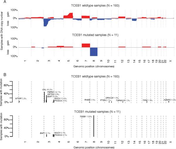
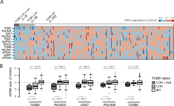
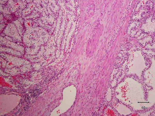
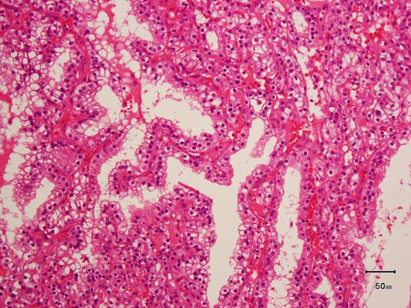
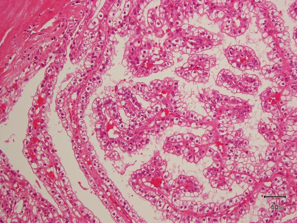
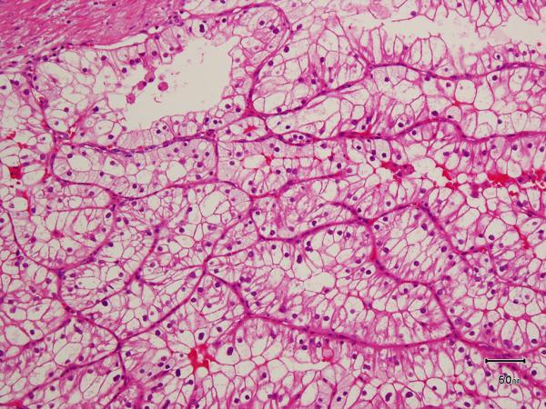
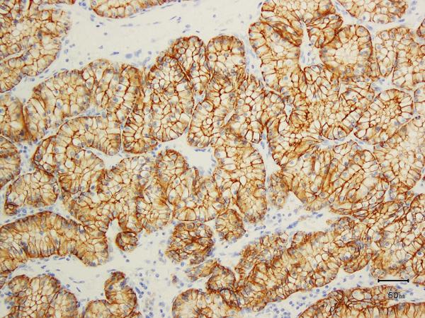
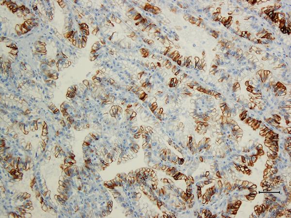
Integrated sequencing analysis identified a group of tumors among clear cell renal cell carcinomas characterized by hotspot mutations in TCEB1 (a gene that contributes to the VHL complex to ubiquitinate hypoxia-inducible factor). We analyzed 11 tumors from two distinct cohorts with TCEB1 mutations along with an expanded cohort to assess whether these should be considered an entity distinct from clear cell renal cell carcinoma and clear cell papillary renal cell carcinoma. All tumors were characterized by hotspot mutations in TCEB1 Y79C/S/F/N or A100P. Morphological and immunohistochemical characteristics of the tumors were assessed by two experienced genitourinary pathologists. Clinical and pathological variables, copy number alterations, mutations, and expression signatures were compared with a cohort of TCEB1 wild-type tumors. All TCEB1-mutated tumors were VHL and PBRM1 wild type and contained distinct copy number profiles including loss of heterozygosity of chromosome 8, the location of TCEB1 (8q21.11). All tumors lacked the clear cell renal cell carcinoma signature 3p loss and contained distinct gene expression signatures. None of the clear cell papillary tumors harbored TCEB1 mutations. Pathologically, all TCEB1-mutated tumors shared characteristic features including thick fibromuscular bands transecting the tumor, pure clear cell cytology frequently with cells showing voluminous cytoplasm, and clear cell renal cell carcinoma-like acinar areas associated with infolding tubular and focally papillary architecture. The presence of voluminous cytoplasm, absence of luminal polarization of tumor nuclei, and lack of extensive cup-like distribution of carbonic anhydrase-IX expression distinguish it from clear cell papillary carcinoma. None of the patients developed metastases at last follow-up (median 48 months). In sum, TCEB1-mutated renal cell carcinoma is a distinct entity with recurrent hotspot mutations, specific copy number alterations, pathway activation, and characteristic morphological features. Further clinical follow-up is needed to determine whether these tumors are more indolent compared with the conventional clear cell renal cell carcinoma.


Figures

Figure 1
Figure 1
A) Genome wide copy number plots depicting chromosomal gains (red) and losses (blue) in low grade clear cell renal cell carcinomasamples from the TCGA vs TCEB1 mutated samples in the combined TCGA and Sato et al data sets. B) Recurrent mutations in both the combined low grade clear cell renal cell carcinomacohort vs TCEB1 mutated samples.
Figure 2
Figure 2
Genome wide mRNA expression correlation between Sato et al and TCGA datasets when comparing TCEB1 mutated tumors vs low grade clear cell renal cell carcinoma(X and Y axses respectively).
Figure 3
Figure 3
Pathway analysis reveals down regulation of Pol II Elongation genes in TCEB1 mutated tumors. A) Heatmap of mRNA expression of Pol II elongation genes stratified by TCEB1 mutation and concomitant loss of heterozygosity (loss of heterozygosity ), loss of heterozygosity alone, and in the wildtype setting. B) Box plot of specific Pol II elongation genes demonstrating a lack of expression changes in tumors with loss of heterozygosity alone compared to combined TCEB1 mutation and loss of heterozygosity .
Figure 4
Figure 4
A) Two nodules of the tumor separated by a thick band of fibromuscular stroma, B) branching tubules in the tumor; the branchings and infoldings give the appearance of papillations, C) long, true papillations are also common in the tumor, D) the clear-appearing cells in the tumor often show fine granules and fibrillations in the cytoplasm; the cells typically appear voluminous with prominent cell membranes, E) immunohistochemical stain for carbonic anhydrase-IX (CA-IX) shows diffuse, membranous (box-like) positivity in all tumors, and F) all tumors are immunoreactive for cytokeratin 7 (CK7), exhibiting patchy (as shown here) to more diffuse positivity.
Figure 4
Figure 4
A) Two nodules of the tumor separated by a thick band of fibromuscular stroma, B) branching tubules in the tumor; the branchings and infoldings give the appearance of papillations, C) long, true papillations are also common in the tumor, D) the clear-appearing cells in the tumor often show fine granules and fibrillations in the cytoplasm; the cells typically appear voluminous with prominent cell membranes, E) immunohistochemical stain for carbonic anhydrase-IX (CA-IX) shows diffuse, membranous (box-like) positivity in all tumors, and F) all tumors are immunoreactive for cytokeratin 7 (CK7), exhibiting patchy (as shown here) to more diffuse positivity.

References

    1. Srigley JR, Delahunt B, Eble JN, et al. The International Society of Urological Pathology (ISUP) Vancouver Classification of Renal Neoplasia. Am J Surg Pathol. 2013;37:1469–89. - PubMed
    1. Rohan SM, Xiao Y, Liang Y, et al. Clear-cell papillary renal cell carcinoma: molecular and immunohistochemical analysis with emphasis on the von Hippel-Lindau gene and hypoxia-inducible factor pathway-related proteins. Mod Pathol. 2011;24:1207–20. - PubMed
    1. Cancer Genome Atlas Research N, Analysis working group: Baylor College of M. Creighton CJ, et al. Comprehensive molecular characterization of clear cell renal cell carcinoma. Nature. 2013;499:43–9. - PMC - PubMed
    1. Sato Y, Yoshizato T, Shiraishi Y, et al. Integrated molecular analysis of clear-cell renal cell carcinoma. Nat Genet. 2013;45:860–7. - PubMed
    1. Varela I, Tarpey P, Raine K, et al. Exome sequencing identifies frequent mutation of the SWI/SNF complex gene PBRM1 in renal carcinoma. Nature. 2011;469:539–42. - PMC - PubMed

Publication types