. 2015 Apr 15:405:14-24.
doi: 10.1016/j.mce.2015.02.006. Epub 2015 Feb 9.

FOXO1 is regulated by insulin and IGF1 in pituitary gonadotropes

Affiliations

FOXO1 is regulated by insulin and IGF1 in pituitary gonadotropes

Danalea V Skarra et al. Mol Cell Endocrinol. .

Abstract

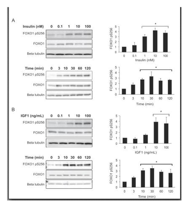
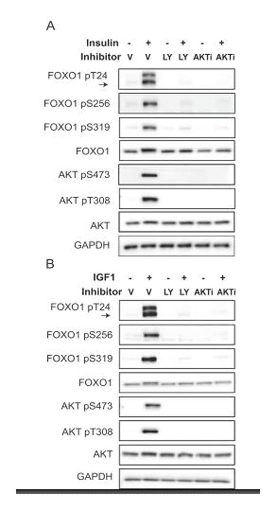
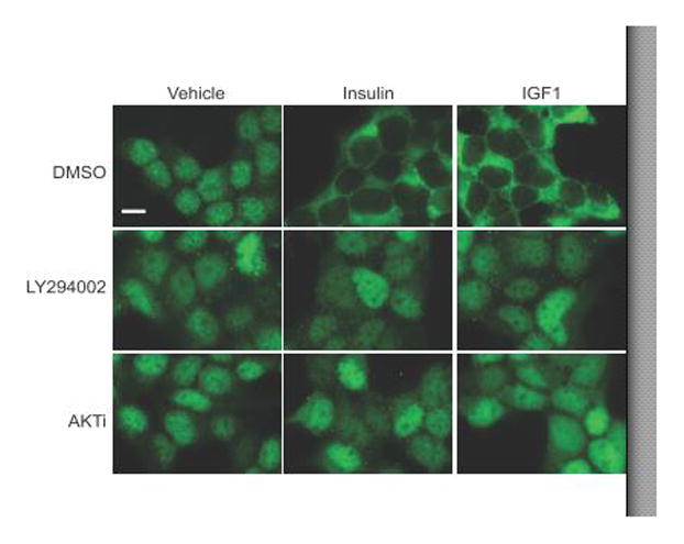
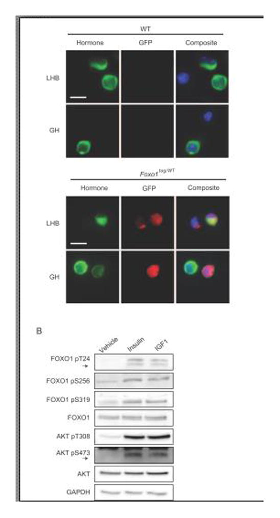
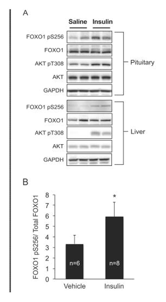
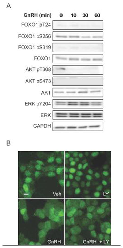
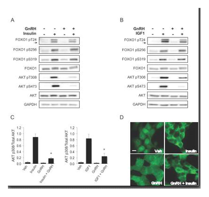
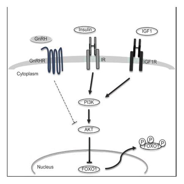
The FOXO1 transcription factor is important for multiple aspects of reproductive function. We previously reported that FOXO1 functions as a repressor of gonadotropin hormone synthesis, but how FOXO1 is regulated in pituitary gonadotropes is unknown. The growth factors, insulin and insulin-like growth factor I (IGF1), function as key regulators of cell proliferation, metabolism and apoptosis in multiple cell types through the PI3K/AKT signaling pathway. In this study, we found that insulin and IGF1 signaling in gonadotropes induced FOXO1 phosphorylation through the PI3K/AKT pathway in immortalized and primary cells, resulting in FOXO1 relocation from the nucleus to the cytoplasm. Furthermore, insulin administration in vivo induced phosphorylation of FOXO1 and AKT in the pituitary. Thus, insulin and IGF1 act as negative regulators of FOXO1 activity and may serve to fine-tune gonadotropin expression.

Keywords: Forkhead box O1; GnRH; Gonadotrope; IGF1; Insulin.


Figures

Figure 1
Figure 1. Insulin and IGF1 induce phosphorylation of FOXO1 Ser256 in immortalized gonadotropes
A. Left: westerns of LβT2 cells that were placed in serum-free media overnight, then treated with increasing concentrations of insulin for 10 min or with 10 nM insulin for up to 2 hrs. Right: FOXO1 pSer256 densitometry values were normalized to total FOXO1, then graphed as fold untreated. B. Left: westerns of LβT2 cells that were placed in serum-free media overnight, then treated with increasing concentrations of IGF1 for 10 min or with 10 ng/mL IGF1 for up to 2 hrs. Right: FOXO1 pSer256 densitometry values were normalized to total FOXO1 and expressed as fold untreated. Graphed data represent mean and error bars as SEM, n=3. For statistical analysis, pSer256/total FOXO1 data were analyzed by randomized block one-way analysis of variance (ANOVA) (82) and post-hoc Dunnett’s test using the statistical package JMP 11.0 (SAS, Cary, NC). Significant differences from control were designated as p<0.05 and represented with an asterisk.
Figure 2
Figure 2. Insulin- and IGF1-induced phosphorylation of FOXO1 at Thr24, Ser256 and Ser319 is dependent on PI3K/AKT signaling in LβT2 cells
Cells were placed in serum-free media overnight. The next day, cells were pretreated with DMSO (Vehicle, V), LY294002 50 μM (LY) or AKTVIII 3 μM (AKTi) for 1 hr and then treated with insulin (10 nM) or IGF1 (10 ng/mL) for 10 min. FOXO1 and AKT phosphorylation changes were assessed by western. Arrow indicates FOXO1 pT24 band; upper band is FOXO3 pT24. Images are representative of 3 independent experiments.
Figure 3
Figure 3. Insulin- and IGF1-stimulated cytoplasmic localization of FOXO1 is dependent on PI3K/AKT signaling
LβT2 cells grown on poly-L-lysine coverslips were placed in serum-free media overnight. The next day, cells were pretreated with DMSO (Vehicle), LY294002 50 μM or AKTVIII 3 μM (AKTi) for 1 hr, then treated with insulin (10 nM) or IGFI (10 ng/mL) for 30 min. Cells were fixed and stained with primary FOXO1 antibody and secondary Alexa Fluor 488 (green) for immunofluorescence microscopy. Images are representative of 3 independent experiments. Scale bar=10 μm.
Figure 4
Figure 4. In cultured murine primary pituitary cells, FOXO1 and AKT are phosphorylated in response to insulin and IGF1
A. Cultured primary pituitary cells from FOXO1tag/WT and WT mice were grown on poly-L-lysine coverslips, fixed and stained with primary GFP antibody along with primary LHB or GH antibody. FOXO1-GFP was detected using secondary Alex Fluor 594 (red), hormones with secondary DyLight 488 (green) and the nuclear compartment with DAPI (blue). Scale bar=10 μm. Images are representative of 3 independent experiments. B. Cultured primary pituitary cells from WT mice were placed in serum-free media for 16 hrs, then treated with insulin (10 nM) or IGF1 (10 ng/mL) for 30 min prior to lysis. Phosphorylation changes in FOXO1 and AKT were assessed by western. Images are representative of 2 independent experiments.
Figure 5
Figure 5. Insulin increases pituitary FOXO1 phosphorylation in vivo
Male mice were fasted for 6 hours, then given either saline or regular insulin (1.5 U/kg) by intraperitoneal injection. After 10 minutes, the mice were sacrificed and their pituitaries and livers harvested and immediately placed in liquid nitrogen. A. Changes in phosphorylation were assessed by western and representative blots are shown for 4 animals, two saline (vehicle) and two insulin. Insulin-induced phosphorylation of FOXO1 Ser256 and AKT Thr308 in the liver were used as positive controls. B. Pituitary FOXO1 pS256 densitometry values were normalized to total FOXO1. Data are presented as the mean and error bars are SEM, n=6 (Vehicle), n=8 (Insulin). The asterisk indicates that insulin significantly increased pituitary FOXO1 Ser256 phosphorylation compared to saline vehicle using Student’s t test, p<0.05.
Figure 6
Figure 6. GnRH does not regulate FOXO1 phosphorylation
A. LβT2 cells were placed in serum-free media overnight and treated the next day with 10 nM GnRH for up to 2 hrs. Changes in FOXO1 and AKT phosphorylation were assessed by western. Images are representative of 3 independent experiments. B. LβT2 cells were placed in serum-free media overnight. The following day, cells were treated with DMSO (Veh) or LY294002 (LY) 50 μM for 1 hr and then GnRH 10 nM was added for 30 min. Cells were fixed and stained with primary FOXO1 antibody and secondary Alexa Fluor 488 (green) for immunofluorescence microscopy. Images are representative of 5 independent experiments. Scale bar=10 μm
Figure 7
Figure 7. GnRH and insulin co-treatment diminishes AKT phosphorylation, but FOXO1 is not affected
A and B. LβT2 cells were placed in serum-free media overnight. The following day, cells were treated with (A) insulin 10 nM or (B) IGF1 10 ng/mL and GnRH 10 nM co-treatment for 30 min and lysed for western analyses. C. AKT p308 densitometry values were normalized to total AKT and graphed as the mean and error bars as SEM, n=3. Significant interaction was designated by an asterisk as defined by two-way ANOVA from insulin or IGF1 alone, p<0.05. D. Immunofluorescence of LβT2 cells under the same treatment conditions as for westerns in A and B above, then fixed and stained with primary FOXO1 antibody and secondary Alexa Fluor 488 (green). Scale bar=10 μm. All images are representative of 3 independent experiments.
Figure 8
Figure 8. Model of growth factor signaling to FOXO1 in pituitary gonadotrope cells
Insulin and IGF1 signal through their cognate receptors to PI3K and AKT, which directly phosphorylates FOXO1. AKT phosphorylation of FOXO1 causes its relocation to the cytoplasm. GnRH receptor activation can also attenuate insulin- or IGF1-induced AKT activity, possibly at the level of AKT, but FOXO1 inhibition by insulin or IGF1 still results in FOXO1 relocation to the cytoplasm. GnRHR, GnRH receptor; IR, insulin receptor; IGF1R, IGF1 receptor.

Similar articles

Cited by

References

    1. Galili N, Davis RJ, Fredericks WJ, Mukhopadhyay S, Rauscher FJ, 3rd, Emanuel BS, Rovera G, Barr FG. Fusion of a fork head domain gene to PAX3 in the solid tumour alveolar rhabdomyosarcoma. Nat Genet. 1993;5:230–235. - PubMed
    1. Nakamura N, Ramaswamy S, Vazquez F, Signoretti S, Loda M, Sellers WR. Forkhead transcription factors are critical effectors of cell death and cell cycle arrest downstream of PTEN. Mol Cell Biol. 2000;20:8969–8982. - PMC - PubMed
    1. Hosaka T, Biggs WH, 3rd, Tieu D, Boyer AD, Varki NM, Cavenee WK, Arden KC. Disruption of forkhead transcription factor (FOXO) family members in mice reveals their functional diversification. Proc Natl Acad Sci USA. 2004;101:2975–2980. - PMC - PubMed
    1. Kousteni S. FoxO1, the transcriptional chief of staff of energy metabolism. Bone. 2012;50:437–443. - PMC - PubMed
    1. Battiprolu PK, Hojayev B, Jiang N, Wang ZV, Luo X, Iglewski M, Shelton JM, Gerard RD, Rothermel BA, Gillette TG, Lavandero S, Hill JA. Metabolic stress–induced activation of FoxO1 triggers diabetic cardiomyopathy in mice. J Clin Invest. 2012;122:1109–1118. - PMC - PubMed

Publication types

Substances