

Electron beam sterilization does not have a detrimental effect on the ability of extracellular matrix scaffolds to support in vivo ligament healing
- PMID: 25676876
- PMCID: PMC4517185
- DOI: 10.1002/jor.22855
Electron beam sterilization does not have a detrimental effect on the ability of extracellular matrix scaffolds to support in vivo ligament healing
Abstract
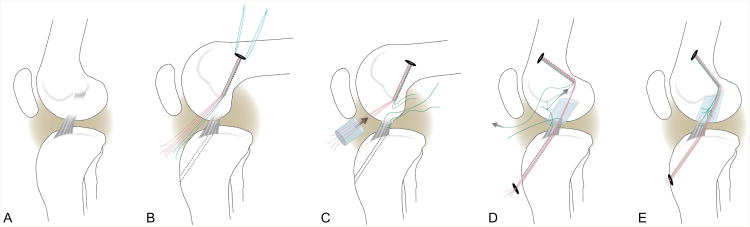
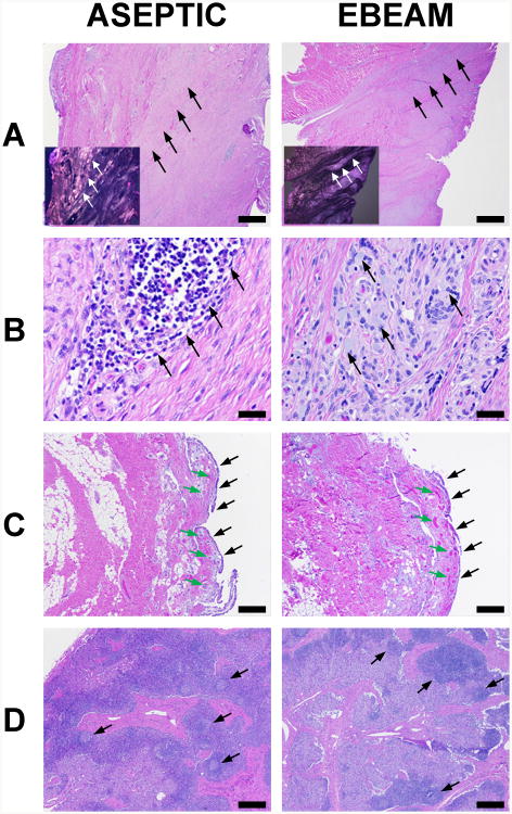
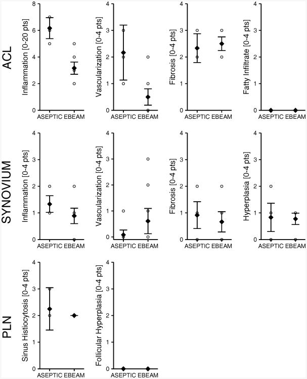
Extracellular matrix (ECM) scaffolds have been used to enhance anterior cruciate ligament (ACL) repair in large animal models. To translate this technology to clinical care, identifying a method which effectively sterilizes the material without significantly impairing in vivo function is desirable. Sixteen Yorkshire pigs underwent ACL transection and were randomly assigned to bridge-enhanced ACL repair-primary suture repair of the ACL with addition of autologous blood soaked ECM scaffold--with either (i) an aseptically processed ECM scaffold, or (ii) an electron beam irradiated ECM scaffold. Primary outcome measures included sterility of the scaffold and biomechanical properties of the scaffold itself and the repaired ligament at 8 weeks after surgery. Scaffolds treated with 15 kGy electron beam irradiation had no bacterial or fungal growth noted, while aseptically processed scaffolds had bacterial growth in all tested samples. The mean biomechanical properties of the scaffold and healing ligament were lower in the electron beam group; however, differences were not statistically significant. Electron beam irradiation was able to effectively sterilize the scaffolds. In addition, this technique had only a minimal impact on the in vivo function of the scaffolds when used for ligament healing in the porcine model.
Keywords: anterior cruciate ligament; bridge-enhanced ACL repair; collagen scaffold; electron beam irradiation.
© 2015 Orthopaedic Research Society. Published by Wiley Periodicals, Inc.
Figures
Similar articles
-
Effect of low-temperature ethylene oxide and electron beam sterilization on the in vitro and in vivo function of reconstituted extracellular matrix-derived scaffolds.J Biomater Appl. 2015 Oct;30(4):435-49. doi: 10.1177/0885328215590967. Epub 2015 Jun 18. J Biomater Appl. 2015. PMID: 26088294 Free PMC article.
-
Biomechanical Outcomes of Bridge-enhanced Anterior Cruciate Ligament Repair Are Influenced by Sex in a Preclinical Model.Clin Orthop Relat Res. 2015 Aug;473(8):2599-608. doi: 10.1007/s11999-015-4226-9. Clin Orthop Relat Res. 2015. PMID: 25742916 Free PMC article.
-
Use of a bioactive scaffold to stimulate anterior cruciate ligament healing also minimizes posttraumatic osteoarthritis after surgery.Am J Sports Med. 2013 Aug;41(8):1762-70. doi: 10.1177/0363546513483446. Epub 2013 Jul 15. Am J Sports Med. 2013. PMID: 23857883 Free PMC article.
-
Addition of autologous mesenchymal stem cells to whole blood for bioenhanced ACL repair has no benefit in the porcine model.Am J Sports Med. 2015 Feb;43(2):320-30. doi: 10.1177/0363546514559826. Epub 2014 Dec 30. Am J Sports Med. 2015. PMID: 25549633 Free PMC article.
-
Anterior Cruciate Ligament: Structure, Injuries and Regenerative Treatments.Adv Exp Med Biol. 2015;881:161-86. doi: 10.1007/978-3-319-22345-2_10. Adv Exp Med Biol. 2015. PMID: 26545750 Review.
Cited by
-
Early MRI-based quantitative outcomes are associated with a positive functional performance trajectory from 6 to 24 months post-ACL surgery.Knee Surg Sports Traumatol Arthrosc. 2023 May;31(5):1690-1698. doi: 10.1007/s00167-022-07000-8. Epub 2022 Jun 15. Knee Surg Sports Traumatol Arthrosc. 2023. PMID: 35704062 Free PMC article. Clinical Trial.
-
Bridge-Enhanced Anterior Cruciate Ligament Repair: Two-Year Results of a First-in-Human Study.Orthop J Sports Med. 2019 Mar 22;7(3):2325967118824356. doi: 10.1177/2325967118824356. eCollection 2019 Mar. Orthop J Sports Med. 2019. PMID: 30923725 Free PMC article.
-
The Bridge-Enhanced Anterior Cruciate Ligament Repair (BEAR) Procedure: An Early Feasibility Cohort Study.Orthop J Sports Med. 2016 Nov 21;4(11):2325967116672176. doi: 10.1177/2325967116672176. eCollection 2016 Nov. Orthop J Sports Med. 2016. PMID: 27900338 Free PMC article.
-
Optimizing outcomes of ACL surgery-Is autograft reconstruction the only reasonable option?J Orthop Res. 2021 Sep;39(9):1843-1850. doi: 10.1002/jor.25128. Epub 2021 Jul 16. J Orthop Res. 2021. PMID: 34191344 Free PMC article. Review.
-
Effect of low-temperature ethylene oxide and electron beam sterilization on the in vitro and in vivo function of reconstituted extracellular matrix-derived scaffolds.J Biomater Appl. 2015 Oct;30(4):435-49. doi: 10.1177/0885328215590967. Epub 2015 Jun 18. J Biomater Appl. 2015. PMID: 26088294 Free PMC article.
References
-
-
11139:2006. IT. 2006. Sterilization of health care products - vocabulary.
-
-
- Favero M. In: Sterility assurance: concepts for patient safety, in disinfection, sterilization and antisepsis: principles and practices in healthcare facilities, chapter 12. WA R, editor. Washington: Association for Professionals in Infection Control and Epidemiology. DC.; 2001. pp. 110–119.
-
-
ANSI/AAMI_ST67:2003. 203. Sterilization of health care products - requirements for products labeled “STERILE”.
-
-
- Hemmerich KJ, et al. Sterilization methods stand the test of time. Medical Device and Diagnostic Industry. 2004;26
-
- Daly KA, Liu S, Agrawal V, et al. Damage associated molecular patterns within xenogeneic biologic scaffolds and their effects on host remodeling. Biomaterials. 2012;33:91–101. - PubMed
Publication types
MeSH terms
Grants and funding
LinkOut - more resources
Full Text Sources
Other Literature Sources
Medical
