

Handheld photoacoustic probe to detect both melanoma depth and volume at high speed in vivo
- PMID: 25676898
- PMCID: PMC4530093
- DOI: 10.1002/jbio.201400143
Handheld photoacoustic probe to detect both melanoma depth and volume at high speed in vivo
Abstract
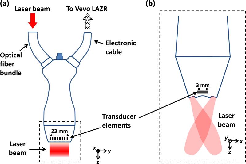
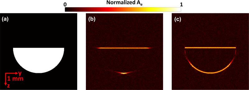
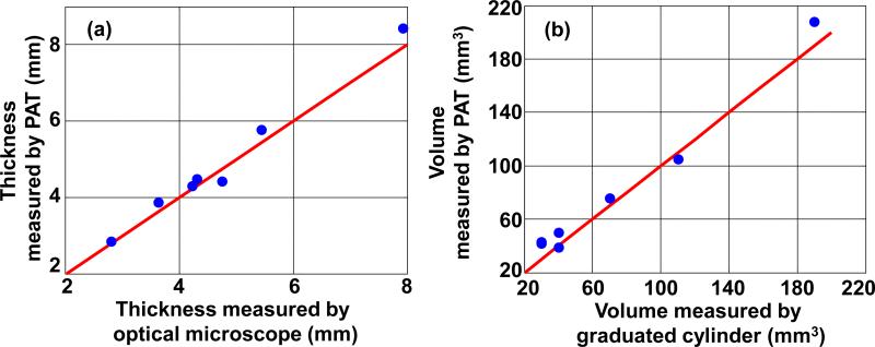
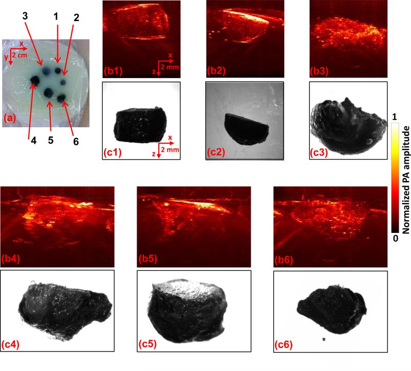
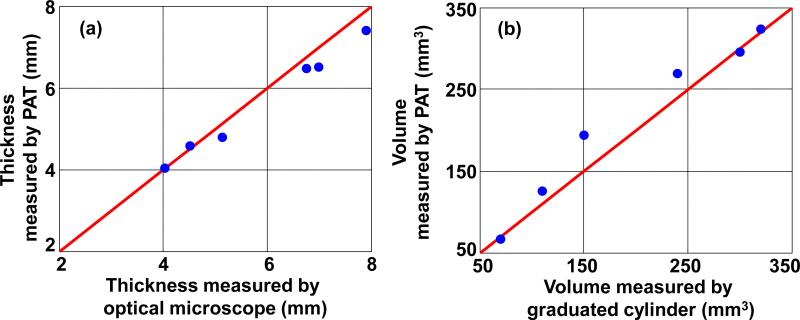
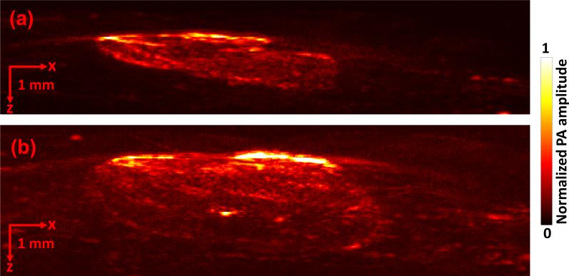
We applied a linear-array-based photoacoustic probe to detect melanin-containing melanoma tumor depth and volume in nude mice in vivo. This system can image melanomas at five frames per second (fps), which is much faster than our previous handheld single transducer system (0.1 fps). We first theoretically show that, in addition to the higher frame rate, almost the entire boundary of the melanoma can be detected by the linear-array-based probe, while only the horizontal boundary could be detected by the previous system. Then we demonstrate the ability of this linear-array-based system in measuring both the depth and volume of melanoma through phantom, ex vivo, and in vivo experiments. The volume detection ability also enables us to accurately calculate the rate of growth of the tumor, which is an important parameter in quantifying the tumor activity. Our results show that this system can be used for clinical melanoma diagnosis and treatment in humans at the bedside. Linear-array-based PA images of melanoma acquired in vivo on day 3 (a) and day 6 (b).
Keywords: handheld; melanoma; photoacoustic; rate of growth.
© 2014 WILEY-VCH Verlag GmbH & Co. KGaA, Weinheim.
Figures
Similar articles
-
Handheld photoacoustic microscopy to detect melanoma depth in vivo.Opt Lett. 2014 Aug 15;39(16):4731-4. doi: 10.1364/OL.39.004731. Opt Lett. 2014. PMID: 25121860 Free PMC article.
-
Non-invasive dynamic assessment of conjunctival melanomas by photoacoustic imaging.Exp Eye Res. 2019 Feb;179:157-167. doi: 10.1016/j.exer.2018.11.014. Epub 2018 Nov 14. Exp Eye Res. 2019. PMID: 30447197
-
Multispectral photoacoustic imaging for the detection of subclinical melanoma.J Surg Oncol. 2019 Jun;119(8):1070-1076. doi: 10.1002/jso.25447. Epub 2019 Mar 15. J Surg Oncol. 2019. PMID: 30874312
-
Real-time photoacoustic tomograpghy using linear array probe and detection of line structure using Hough transform.Biomed Mater Eng. 2015;26 Suppl 1:S1483-90. doi: 10.3233/BME-151447. Biomed Mater Eng. 2015. PMID: 26405912
-
Photoacoustic imaging: a potential tool to detect early indicators of metastasis.Expert Rev Med Devices. 2013 Jan;10(1):125-34. doi: 10.1586/erd.12.62. Expert Rev Med Devices. 2013. PMID: 23278229 Free PMC article. Review.
Cited by
-
Photoacoustic elastography.Opt Lett. 2016 Feb 15;41(4):725-8. doi: 10.1364/OL.41.000725. Opt Lett. 2016. PMID: 26872173 Free PMC article.
-
Bioinspired Multifunctional Melanin-Based Nanoliposome for Photoacoustic/Magnetic Resonance Imaging-Guided Efficient Photothermal Ablation of Cancer.Theranostics. 2018 Feb 7;8(6):1591-1606. doi: 10.7150/thno.22430. eCollection 2018. Theranostics. 2018. PMID: 29556343 Free PMC article.
-
Review of Non-Invasive Imaging Technologies for Cutaneous Melanoma.Biosensors (Basel). 2025 May 7;15(5):297. doi: 10.3390/bios15050297. Biosensors (Basel). 2025. PMID: 40422036 Free PMC article. Review.
-
Photoacoustic imaging for cutaneous melanoma assessment: a comprehensive review.J Biomed Opt. 2024 Jan;29(Suppl 1):S11518. doi: 10.1117/1.JBO.29.S1.S11518. Epub 2024 Jan 12. J Biomed Opt. 2024. PMID: 38223680 Free PMC article.
-
Bond-selective photoacoustic imaging by converting molecular vibration into acoustic waves.Photoacoustics. 2016 Feb 1;4(1):11-21. doi: 10.1016/j.pacs.2016.01.002. eCollection 2016 Mar. Photoacoustics. 2016. PMID: 27069873 Free PMC article. Review.
References
-
- Jemal A, Devesa SS, Hartge P, Tucker MA. Recent trends in cutaneous melanoma incidence among whites in the United States. J. Natl. Cancer Inst. 2001;93(9):678–683. - PubMed
-
- American Cancer Society. http://www.cancer.org/
-
- Ng JC, Swain S, Dowling JP, Wolfe R, Simpson P, Kelly JW. The impact of partial biopsy on histopathologic diagnosis of cutaneous melanoma: experience of an Australian tertiary regerral service. Arch. Dermatol. 2010;146(3):234–239. - PubMed
-
- Ng PC, Barzilai DA, Ismail SA, Averitte RL, Jr., Gilliam AC. Eveluating invasive cutaneous melanoma: is the initial biopsy representative of the final depth? J. Am. Acad. Dermatol. 2003;48(3):420–424. - PubMed
-
- Sellheyer K, Nelson P, Bergfeld WF. Inadequate biopsy technique and specimen size: an alarming tread that compromises patient care and an appeal to our clinical colleagues. Arch. Dermatol. 2010;146(10):1180–1181. - PubMed
Publication types
MeSH terms
Substances
Grants and funding
LinkOut - more resources
Full Text Sources
Other Literature Sources
Medical
Miscellaneous
