

Revascularization of chronic hibernating myocardium stimulates myocyte proliferation and partially reverses chronic adaptations to ischemia
- PMID: 25677430
- PMCID: PMC4328140
- DOI: 10.1016/j.jacc.2014.11.040
Revascularization of chronic hibernating myocardium stimulates myocyte proliferation and partially reverses chronic adaptations to ischemia
Abstract
Background: The time course and extent of recovery after revascularization of viable dysfunctional myocardium are variable. Although fibrosis is a major determinant, myocyte structural and molecular remodeling may also play important roles.
Objectives: This study sought to determine whether persistent myocyte loss and/or irreversibility of protein changes that develop in hibernating myocardium have an impact on functional recovery in the absence of infarction.
Methods: Swine implanted with a chronic left anterior descending artery (LAD) stenosis to produce hibernating myocardium underwent percutaneous revascularization, with serial functional recovery evaluated for 1 month (n = 12). Myocardial tissue was evaluated to assess myocyte size, nuclear density, and proliferation indexes in comparison with those of normal animals and nonrevascularized controls. Proteomic analysis by 2-dimensional differential in-gel electrophoresis was used to determine the reversibility of molecular adaptations of hibernating myocytes.
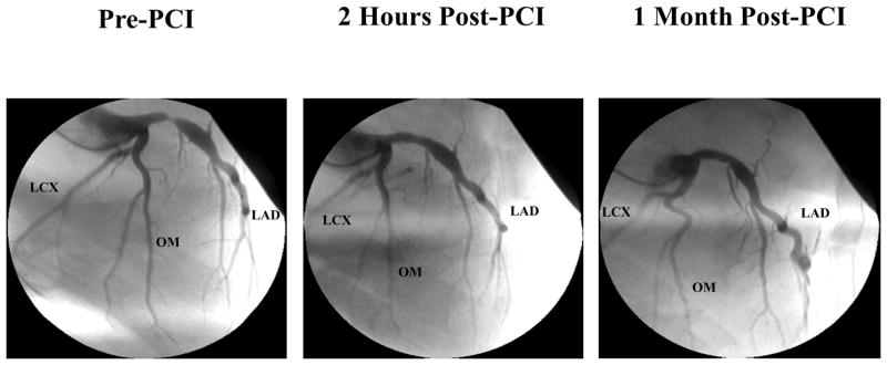
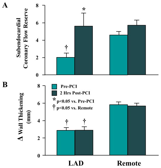
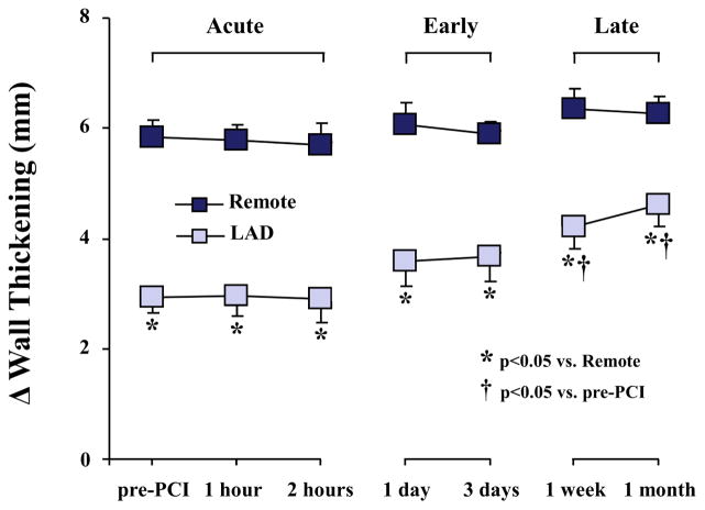
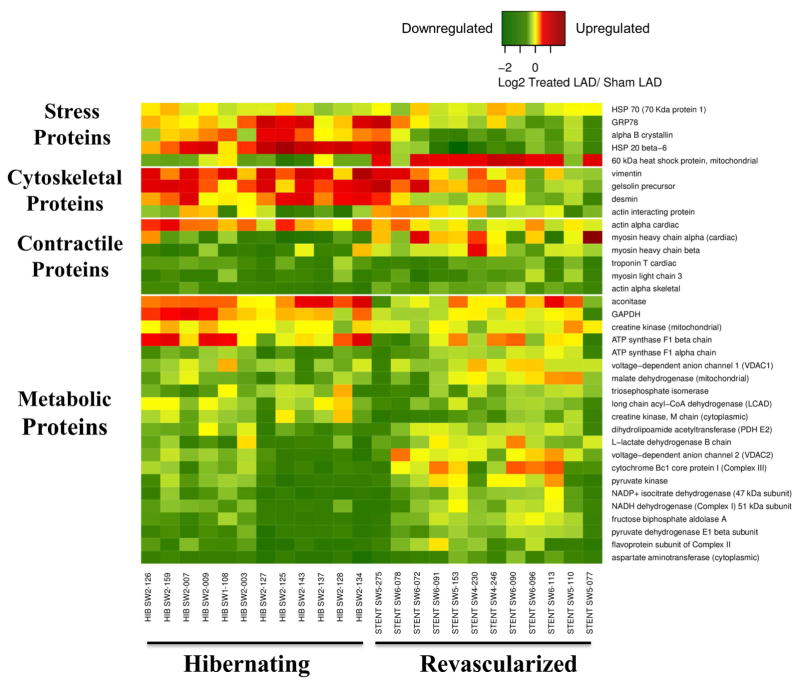
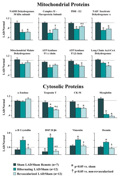
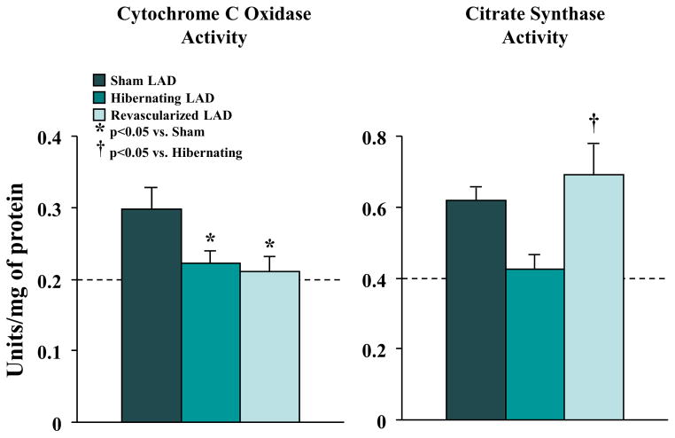
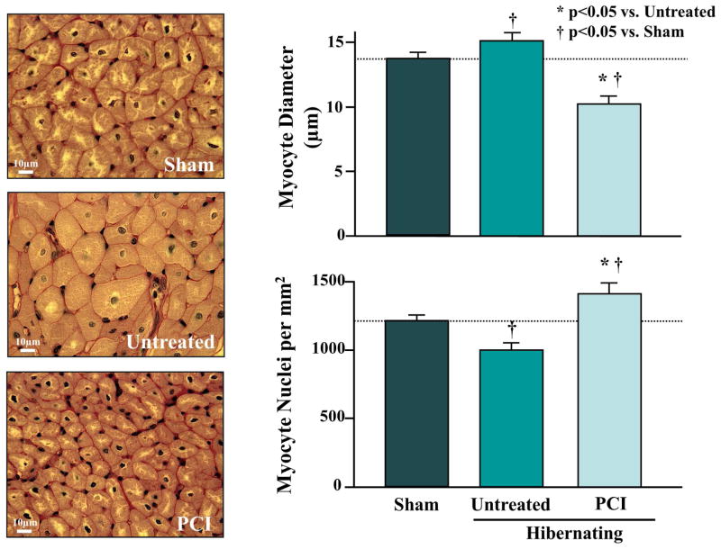
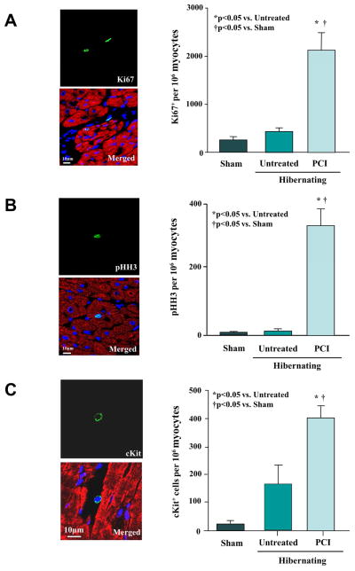
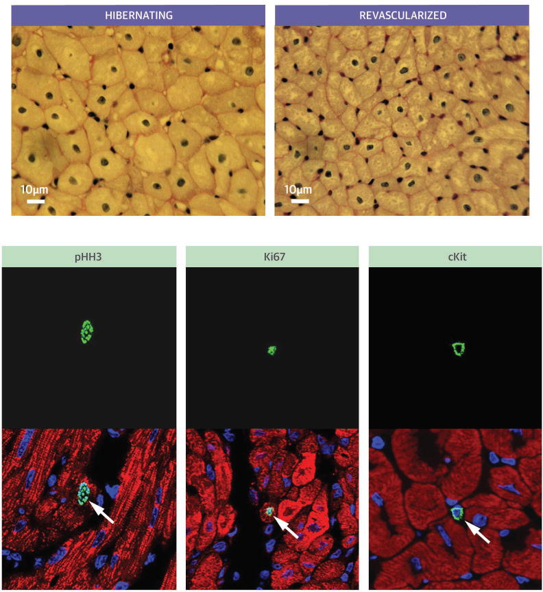
Results: At 3 months, physiological features of hibernating myocardium were confirmed, with depressed LAD wall thickening and no significant infarction. Revascularization normalized LAD flow reserve, with no immediate change in LAD wall thickening. Regional LAD wall thickening slowly improved but remained depressed 1 month post-percutaneous coronary intervention. Surprisingly, revascularization was associated with histological evidence of myocytes re-entering the growth phase of the cell cycle and increases in the number of c-Kit(+) cells. Myocyte nuclear density returned to normal, whereas regional myocyte hypertrophy regressed. Proteomic analysis demonstrated heterogeneous effects of revascularization. Up-regulated stress and cytoskeletal proteins normalized, whereas reduced contractile and metabolic proteins persisted.
Conclusions: Delayed recovery of hibernating myocardium in the absence of scar may reflect persistent reductions in the amounts of contractile and metabolic proteins. Although revascularization appeared to stimulate myocyte proliferation, the persistence of small immature myocytes may have contributed to delayed functional recovery.
Keywords: coronary blood flow; myocyte regeneration; proteomics.
Copyright © 2015 American College of Cardiology Foundation. Published by Elsevier Inc. All rights reserved.
Figures
Comment in
-
Revascularization of hibernating myocardium: uneven reflorescence after the drought.J Am Coll Cardiol. 2015 Feb 24;65(7):698-700. doi: 10.1016/j.jacc.2014.12.024. J Am Coll Cardiol. 2015. PMID: 25677431 No abstract available.
References
-
- Fallavollita JA, Perry BJ, Canty JM., Jr 18F-2-deoxyglucose deposition and regional flow in pigs with chronically dysfunctional myocardium: Evidence for transmural variations in chronic hibernating myocardium. Circulation. 1997;95:1900–9. - PubMed
-
- Vanoverschelde JLJ, Wijns W, Depre C, et al. Mechanisms of chronic regional postischemic dysfunction in humans. New insights from the study of noninfarcted collateral-dependent myocardium. Circulation. 1993;87:1513–23. - PubMed
-
- Lim H, Fallavollita JA, Hard R, et al. Profound apoptosis-mediated regional myocyte loss and compensatory hypertrophy in pigs with hibernating myocardium. Circulation. 1999;100:2380–6. - PubMed
-
- Fallavollita JA, Malm BJ, Canty JM., Jr Hibernating myocardium retains metabolic and contractile reserve despite regional reductions in flow, function, and oxygen consumption at rest. Circ Res. 2003;92:48–55. - PubMed
-
- Page B, Young R, Iyer V, et al. Persistent regional downregulation in mitochondrial enzymes and upregulation of stress proteins in swine with chronic hibernating myocardium. Circ Res. 2008;102:103–12. - PubMed
Publication types
MeSH terms
Substances
Grants and funding
LinkOut - more resources
Full Text Sources
Other Literature Sources
