

Novel method of cell line establishment utilizing fluorescence-activated cell sorting resulting in 6 new head and neck squamous cell carcinoma lines
- PMID: 25677579
- PMCID: PMC4530106
- DOI: 10.1002/hed.24019
Novel method of cell line establishment utilizing fluorescence-activated cell sorting resulting in 6 new head and neck squamous cell carcinoma lines
Abstract
Background: The purpose of this study was to present the establishment of new cell lines, which is important to cancer research.
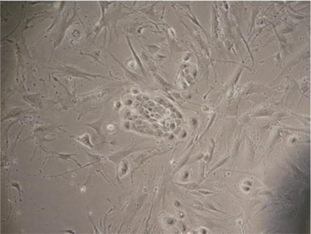
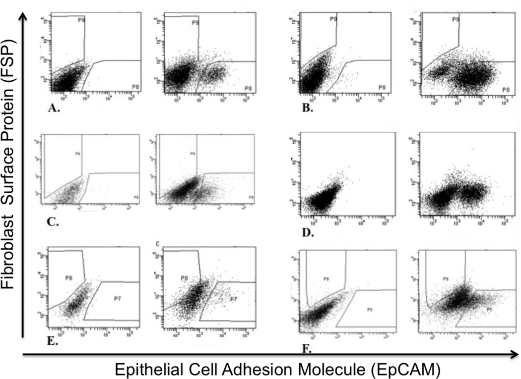
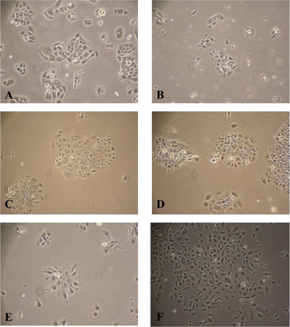
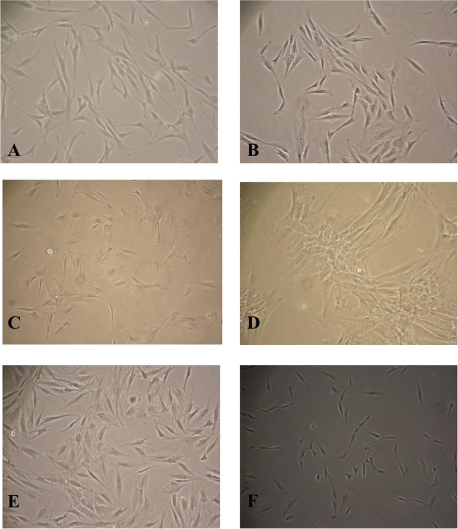
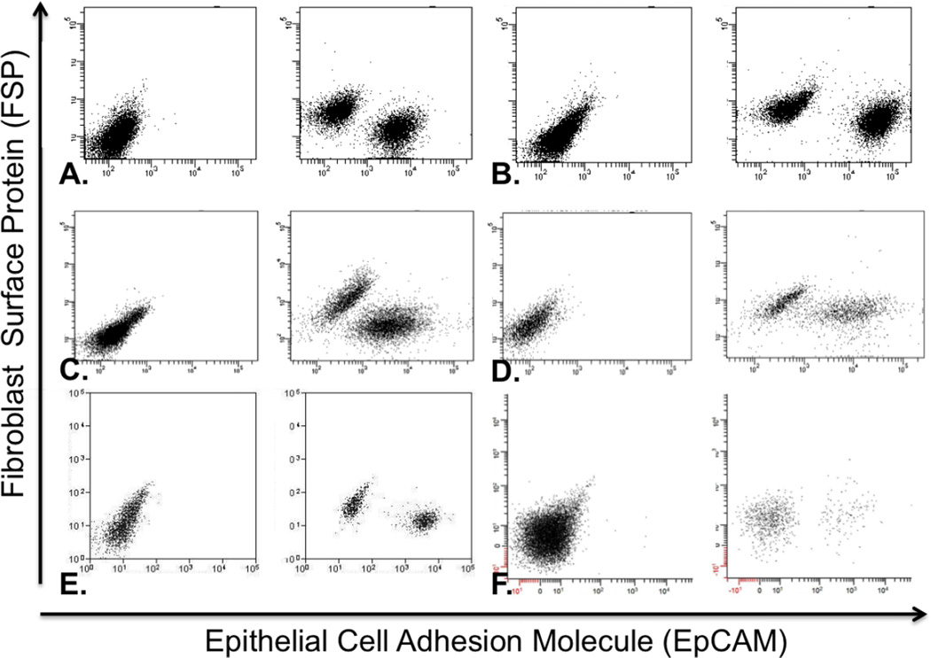
Methods: Six new head and neck squamous cell carcinoma cell lines were established using a novel fluorescence-activated cell sorting (FACS) method in order to overcome the barrier of fibroblast overgrowth and the susceptibility of primary tumors to fail in vitro.
Results: Antibodies chosen for specific targeting of epithelial cells and fibroblasts successfully separated cells for line establishment in 6 of 12 attempts, providing an alternative method of establishing head and neck squamous cell carcinoma cell lines. Each attempt at cell line establishment resulted in an epithelial carcinoma population, which was genotyped and catalogued as a unique cell line, and a corresponding fibroblast population.
Conclusion: The selection of antibody markers could be optimized to aid in the establishment of any cancer cell line derived from any tumor tissue; this method is not limited to head and neck cancer. © 2015 Wiley Periodicals, Inc. Head Neck 38: E459-E467, 2016.
Keywords: cell line; fibroblasts; flow cytometry; squamous cell carcinoma.
© 2015 Wiley Periodicals, Inc.
Figures
References
-
- Harrison RG. Observations on the living developing nerve fiber. Proc. Soc. Exp. Biol. Med. 1907;4:140–143.
-
- Burrows MT. The cultivation of tissues of the chick embryo outside the body. J. Am. Med. Assoc. 1910;55:2057–2058.
-
- Earle WR. Production of malignancy in vitro. IV. The mouse fibroblast cultures and changes seen in living cells. J. Natl Cancer Inst. 1943;4:165–212.
-
- Gey GO, Coffman WD, Kubicek MT. Tissue culture studies of the proliferative capacity of cervical carcinoma and normal epithelium. Cancer Res. 1952;12:264–265.
MeSH terms
Substances
Grants and funding
LinkOut - more resources
Full Text Sources
Other Literature Sources
Medical
Research Materials
