. 2015 Feb 13:5:8454.
doi: 10.1038/srep08454.

How deeply does your mutant sleep? Probing arousal to better understand sleep defects in Drosophila

Affiliations

How deeply does your mutant sleep? Probing arousal to better understand sleep defects in Drosophila

R Faville et al. Sci Rep. .

Abstract

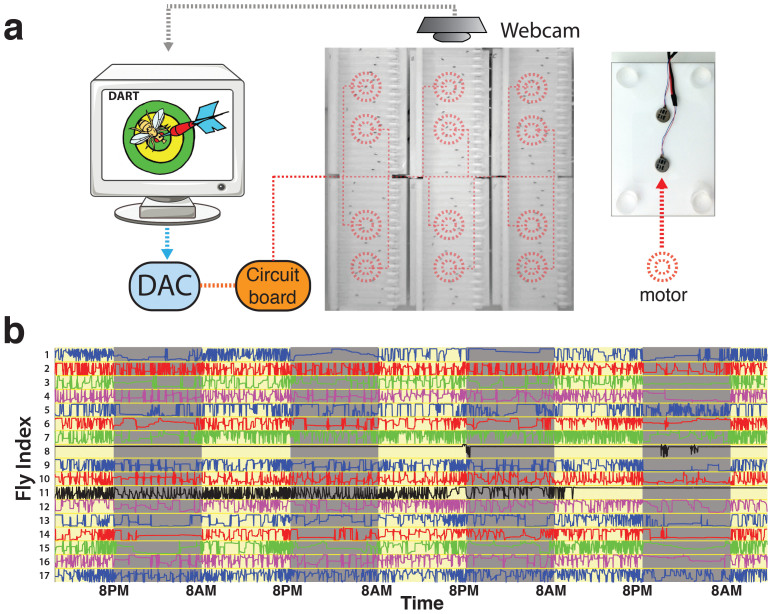
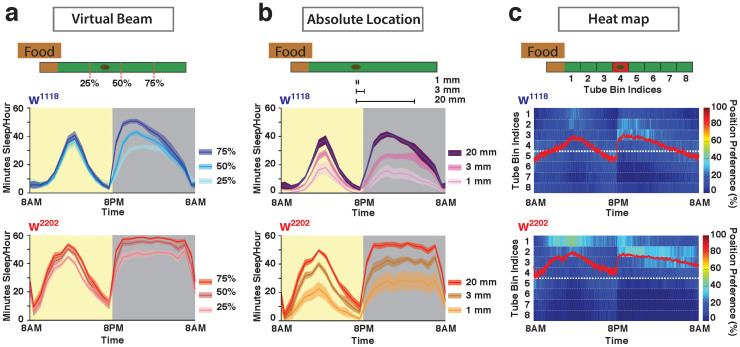
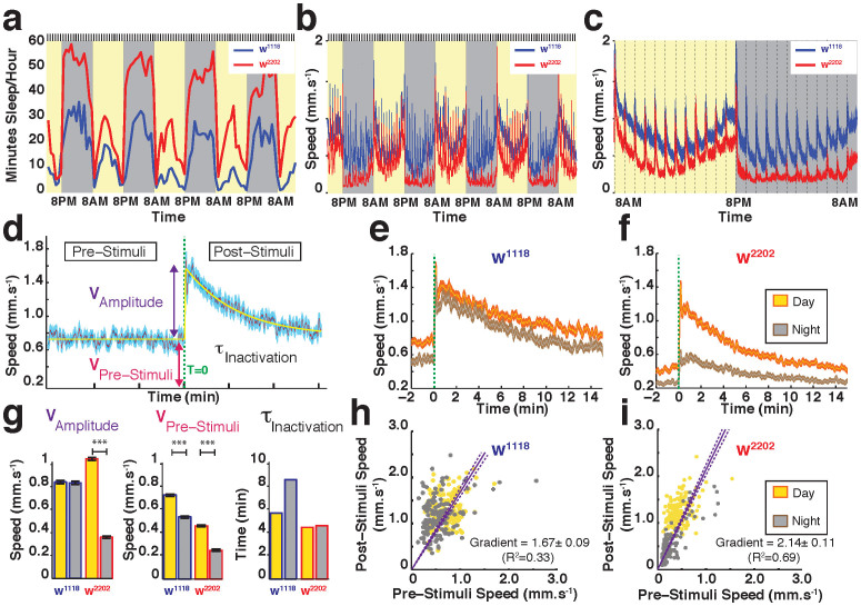
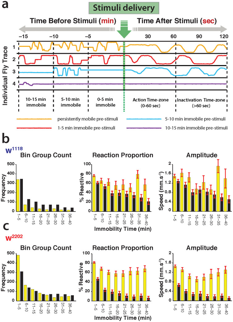
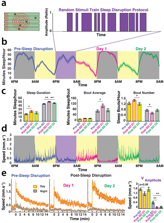
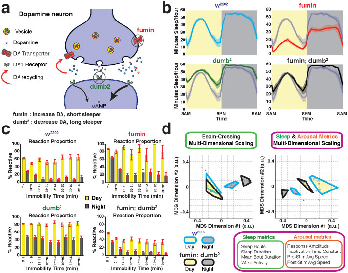
The fruitfly, Drosophila melanogaster, has become a critical model system for investigating sleep functions. Most studies use duration of inactivity to measure sleep. However, a defining criterion for sleep is decreased behavioral responsiveness to stimuli. Here we introduce the Drosophila ARousal Tracking system (DART), an integrated platform for efficiently tracking and probing arousal levels in animals. This video-based platform delivers positional and locomotion data, behavioral responsiveness to stimuli, sleep intensity measures, and homeostatic regulation effects - all in one combined system. We show how insight into dynamically changing arousal thresholds is crucial for any sleep study in flies. We first find that arousal probing uncovers different sleep intensity profiles among related genetic background strains previously assumed to have equivalent sleep patterns. We then show how sleep duration and sleep intensity can be uncoupled, with distinct manipulations of dopamine function producing opposite effects on sleep duration but similar sleep intensity defects. We conclude by providing a multi-dimensional assessment of combined arousal and locomotion metrics in the mutant and background strains. Our approach opens the door for deeper insights into mechanisms of sleep regulation and provides a new method for investigating the role of different genetic manipulations in controlling sleep and arousal.


Figures

Figure 1
Figure 1. The Drosophila ARousal Tracking (DART) system.
(a). Left: DART software records movies via a webcam while simultaneously controlling motor stimuli via a digital to analog converter (DAC). The multiple motors (represented by red dashed circles) are connected to the DAC via a simple circuit board. Middle: Six platforms are positioned in a 3 × 2 arrangement of 17 flies per platform, with two motors per platform. Right: Each platform has two motors glued underneath. The platform has four pedestals (clear circles) that slot into a larger base. (b). Movement traces for 17 flies over four days (yellow background) and nights (grey) outputted from DART. X-axis is time of day and y-axis is horizontal displacement in the tube for each fly. The different colors (blue, red, green purple) are assigned so that the individual traces are easier to distinguish. Graphical user interface supervision allows selection of unusable flies: black traces (flies 8 and 11) have been marked for removal, as flies appeared to have died. Artwork: Benjamin Kottler and Bruno van Swinderen.
Figure 2
Figure 2. Distinct sleep profiles in two related white background strains, w1118 and w2202.
(a). Classical sleep profiles derived from virtual beam-crossing experiments. Flies were tracked continuously, but a virtual “beam” was positioned in three different positions along the 65 mm tubes housing individual flies (25%, 50%, and 75%) to simulate activity readouts from infrared-based tracking systems. Sleep was defined as 5 min or more of inactivity (no beam crossing1). The approximate size of the fly is indicated by a dark oval in the tube. (b). Sleep profiles derived from absolute location data, for both white strains. Sleep was defined as 5 min or more without movement, which was established for 3 different movement thresholds (1 mm, 3 mm, and 20 mm). The 20 mm movement threshold is essentially equivalent in distance to a beam-crossing experiment, as in a. (c). Heat map of position preferences in the tubes for both white strains over multiple days and nights. The individual fly tubes were divided into 8 bins, and position preference (% per unit time over all bins) was plotted as a heat map, with warmer colors indicating a higher probability of flies being in that position. Red line indicates average position for the strain. The thicker white dashed line indicates the mid-point in the tube. See Fig. S2 for additional information on sleep metrics in these strains. N = 45 for w1118 and N = 46 for w2202. Confidence intervals are SEM.
Figure 3
Figure 3. Hourly arousal probing reveals significant differences between white strains.
(a). Average sleep profiles over several days (yellow) and nights (grey) for w1118 (blue) and w2202 (red). Minutes of sleep per hour were determined by absolute location data, based on a 3 mm movement threshold as in Fig. 2b. All flies were stimulated hourly (by vibrations, indicated by the black lines above the graph). N = 47 for w1118 and N = 50 for w2202. (b). Average speed of w1118 (blue) and w2202 (red) for the same set of flies, over the same time period as in a. The responses to the vibrations (black lines above the graph) are evident as spikes in the speed. Data are averaged for all flies, not only immobile ones. (c). Average speed data for day and night for the same set of flies. The vertical dashed lines indicate hourly vibrations testing for behavioral responsiveness. (d). Characterization of behavioral responsiveness curve. Average speed (purple) (±SEM, light blue) is shown for a wild-type strain, with four metrics derived from a best fit (yellow trace) of the average response. Fitted parameters were: the average speed 1 min before the stimulus (or the baseline speed (VPre-Stimuli), the average speed 1 min after the stimulus (Post-Stimuli Speed), the amplitude of the response (VAmplitude), and the time constant of the response (τInactivation). Green dashed line indicates timing of vibration stimulus (T = 0). Flies analyzed for responsiveness profiles were not necessarily immobile. (e). Average response (±SEM) for w1118, for day (yellow) and night (grey). (f). Average response (±SEM) for w2202, for day (yellow) and night (grey). (g). Best fitting response parameters (as outlined in d) for w1118 and w2202 during the day (yellow) and night (grey). N = 2256 responses for w1118 and N = 2400 responses for w2202, divided equally among 47 and 50 flies, respectively, for day and night. (h). Scatterplot of Pre-Stimuli speed versus Post-Stimuli speed, for individual day (yellow dots) and night (grey dots) events in w1118. A linear regression of the data (±SEE) is shown, with associated slope and correlation indicated. (i). Scatterplot of Pre-Stimuli speed versus Post-Stimuli speed, for individual day (yellow dots) and night (grey dots) events in w2202. A regression of the data (±SEE) is shown, with associated slope and correlation indicated.
Figure 4
Figure 4. Sleep intensity differs between the white strains.
(a). Responses were binned according to the duration of prior immobility. Vibration stimuli were delivered once an hour over several days and nights (green arrow). Prior immobility epochs were automatically classified into 5 min bins, as determined by the first retroactive detection of any movement, based on a 3 mm threshold. Four examples are shown, where flies had been immobile for different lengths of time prior to the stimulus (red, blue, and purple lines), and one fly that was moving immediately before the stimulus (gold line). Only immobile flies were included in the subsequent sleep intensity analyses, which summed locomotion responses 1 min after the stimulus (Action time zone). The y-axis represents horizontal fly movement in the tubes. Note that time before and after the stimulus is not on the same scale in the schema. (b). Sleep intensity in w1118. Left panel: frequency count for every 5 min immobility bin, for day (yellow) and night (black). Middle panel: the proportion of flies responding (±SEM) for each immobility bin, for day and night. Right panel: the average amplitude of the response for each immobility bin (average speed ± SEM), for day and night. (c). The same sleep intensity calculations were performed for w2202 N = 47 for w1118 and N = 50 for w2202. See Tables S1–4 for corresponding statistics.
Figure 5
Figure 5. Sleep rebound following a random stimulation protocol.
(a). Random patterns of vibration stimuli (set at 1.2 g) were delivered for 12 hours of night. (b). Average hourly sleep duration (±SEM) in w2202 before (blue), during (purple box), and after (magenta and green) the random stimulation. Yellow background indicates day, grey is night. (c). Traditional sleep duration metrics (±SEM) for the data from b, in the same color scheme. *, p < 0.05; **, P < 0.01, by pair-wise 2-tailed t-test. (d). Average speed (±SEM) for the same flies as in b. (e). Average daytime and nighttime behavioral responsiveness (±SEM) for baseline and two successive days following the random stimulation. Velocity data (Vamplitude) are summarized in the panel on the right, for the same three days. **, P < 0.01, by pair-wise 2-tailed t-test. Yellow plots represent daytime responses whereas grey plots are nighttime responses; blue surrounding indicates the day before the random stimulation, magenta surrounding is one day after the random stimulation, and green surrounding is two days after random stimulation. N = 17 flies.
Figure 6
Figure 6. Sleep and arousal measures can be dissociated with DART.
(a). Opposing effects of fumin and dumb2 on dopamine (DA) function and sleep. Released DA impacts DA1 receptors to initiate cAMP signaling pathways (left black arrow) before being recycled back into the cell (red arrow). fumin is mutant for the DA transporter, leading to increased DA levels in the synapse, and consequently increases cAMP signaling in post-synaptic neurons (right thick black arrow). dumb2 is mutant for the DA1 receptor, leading to decreased cAMP signaling (dashed black arrow). (b). Classical beam-crossing sleep profiles (min sleep/hour (±SEM)) based on a 5 min inactivity criterion, for the w2202 background strain (blue), fumin (red), dumb2 (green), and the double mutant fumin; dumb2 (black). N = 68 for w2202; N = 67 for fumin; N = 62 for dumb2; N = 66 for fumin;dumb2. The w2202 profile is shown in grey for comparison in the three mutant panels. (c). Sleep intensity profiles (% reactive ± SEM) for the same four strains as in b. (d). Multidimensional scaling (MDS) was used to project the data into a two-dimensional space for easier visualization of the multidimensional relationships between different strains. MDS analyses were performed for fumin; dumb2 (blue border) compared to its genetic background strain, w2202 (black border), for daytime (yellow) and nighttime (grey) metrics. Left panel: four beam-crossing metrics (as for b) were used in combination for MDS comparing both strains. The different metrics used are indicated in the green box (bottom of the panel). The daytime effects overlap while the nighttime effects are distinct. Right panel: four arousal-based metrics were combined with four beam-crossing sleep metrics (indicated by the larger magenta box) for MDS analyses, resulting in a complete separation between day and night effects in both strains. a.u., arbitrary units.

Similar articles

Cited by

References

    1. Shaw P. J., Cirelli C., Greenspan R. J. & Tononi G. Correlates of sleep and waking in Drosophila melanogaster. Science 287, 1834–1837 (2000). - PubMed
    1. Hendricks J. C. et al. Rest in Drosophila is a sleep-like state. Neuron 25, 129–138 (2000). - PubMed
    1. Griffith L. C. Neuromodulatory control of sleep in Drosophila melanogaster: integration of competing and complementary behaviors. Current opinion in neurobiology 23, 819–823 (2013). - PMC - PubMed
    1. Tononi G. & Cirelli C. Sleep and synaptic homeostasis: a hypothesis. Brain Res Bull 62, 143–150 (2003). - PubMed
    1. Donlea J. M., Ramanan N. & Shaw P. J. Use-dependent plasticity in clock neurons regulates sleep need in Drosophila. Science 324, 105–108 (2009). - PMC - PubMed

Publication types

LinkOut - more resources