. 2015;37(2):115-30.
doi: 10.1159/000369091. Epub 2015 Feb 10.

The subventricular zone in the immature piglet brain: anatomy and exodus of neuroblasts into white matter after traumatic brain injury

Affiliations

The subventricular zone in the immature piglet brain: anatomy and exodus of neuroblasts into white matter after traumatic brain injury

Beth A Costine et al. Dev Neurosci. 2015.

Abstract

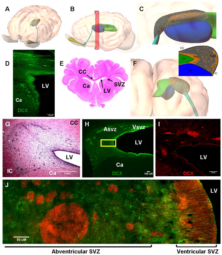
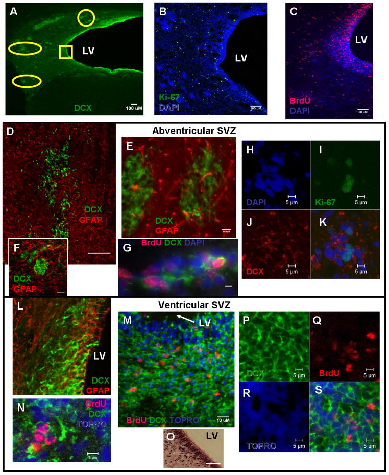
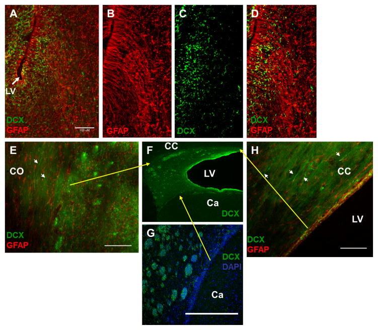
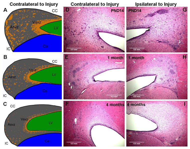
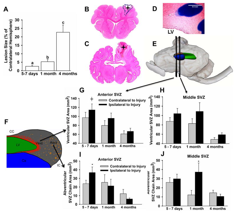
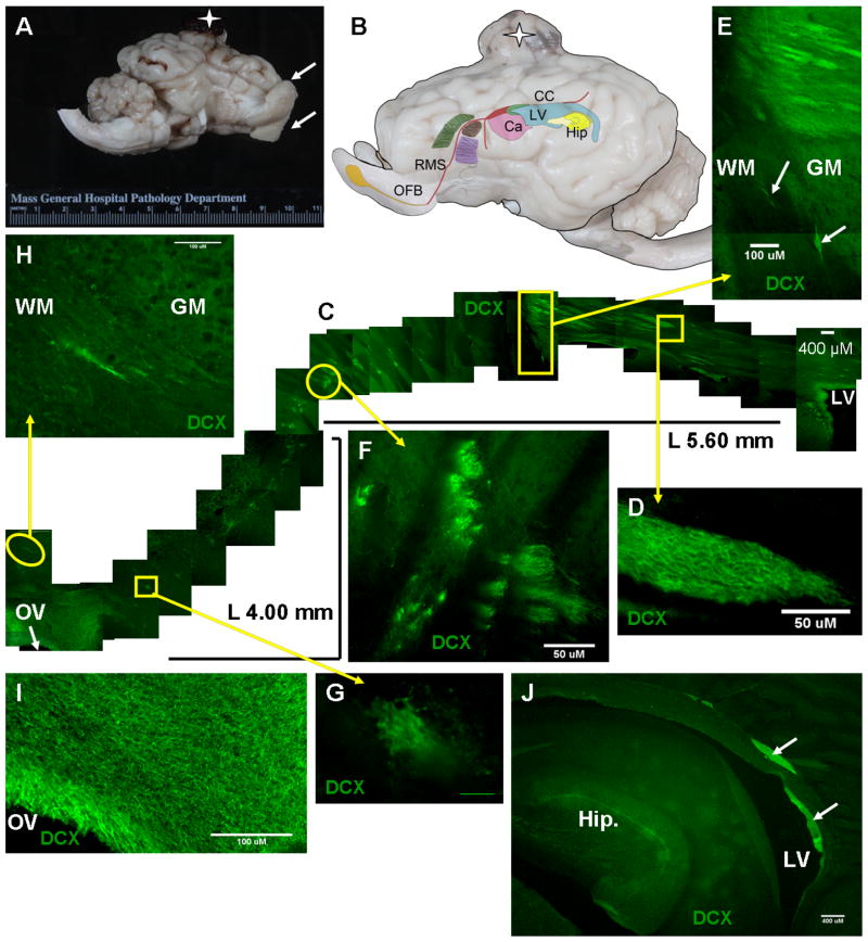
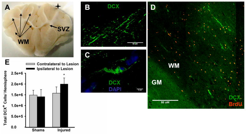
Stimulation of postnatal neurogenesis in the subventricular zone (SVZ) and robust migration of neuroblasts to the lesion site in response to traumatic brain injury (TBI) is well established in rodent species; however, it is not yet known whether postnatal neurogenesis plays a role in repair after TBI in gyrencephalic species. Here we describe the anatomy of the SVZ in the piglet for the first time and initiate an investigation into the effect of TBI on the SVZ architecture and the number of neuroblasts in the white matter. Among all ages of immaturity examined the SVZ contained a dense mesh network of neurogenic precursor cells (doublecortin+) positioned directly adjacent to the ependymal cells (ventricular SVZ, Vsvz) and neuroblasts organized into chains that were distinct from the Vsvz (abventricular SVZ, Asvz). Though the architecture of the SVZ was similar among ages, the areas of Vsvz and Asvz neuroblast chains declined with age. At postnatal day (PND) 14 the white matter tracts have a tremendous number of individual neuroblasts. In our scaled cortical impact model, lesion size increased with age. Similarly, the response of the SVZ to injury was also age dependent. The younger age groups that sustained the proportionately smallest lesions had the largest SVZ areas, which further increased in response to injury. In piglets that were injured at 4 months of age and had the largest lesions, the SVZ did not increase in response to injury. Similar to humans, swine have abundant gyri and gyral white matter, providing a unique platform to study neuroblasts potentially migrating from the SVZ to the lesioned cortex along these white matter tracts. In piglets injured at PND 7, TBI did not increase the total number of neuroblasts in the white matter compared to uninjured piglets, but redistribution occurred with a greater number of neuroblasts in the white matter of the hemisphere ipsilateral to the injury compared to the contralateral hemisphere. At 7 days after injury, less than 1% of neuroblasts in the white matter were born in the 2 days following injury. These data show that the SVZ in the piglet shares many anatomical similarities with the SVZ in the human infant, and that TBI had only modest effects on the SVZ and the number of neuroblasts in the white matter. Piglets at an equivalent developmental stage to human infants were equipped with the largest SVZ and a tremendous number of neuroblasts in the white matter, which may be sufficient in lesion repair without the dramatic stimulation of neurogenic machinery. It has yet to be determined whether neurogenesis and migrating neuroblasts play a role in repair after TBI and/or whether an alteration of normal migration during active postnatal population of brain regions is beneficial in species with gyrencephalic brains.


Figures

Figure 1
Figure 1. Architecture of the uninjured PND 14 piglet subventricular zone and comparison to the human infant
A–B, 3D illustrations of the subventricular zone along the anterior lateral ventricle (green) in the piglet indicating location of coronal sections in E, G, H (green = lateral ventricle, blue = caudate, orange = ventricular SVZ, gray = abventricular SVZ, teal= RMS, yellow = olfactory ventricle). C, Illustration of a sagittal view of the anterior SVZ corresponding to sagittal section in D. The piglet SVZ is a distinct anatomical feature containing a glia (GFAP+) and neuroblasts (DCX+, marker of immature migrating neuroblasts; D, H, J) that correspond to the hematoxylin staining (G). F, 3D illustration of the coronal view of the SVZ and the 2D inset illustrates that the SVZ around the anterior lateral ventricle (LV, green) is surrounded by the corpus callosum (CC) dorsally, the head of caudate (Ca, blue) ventrally, and the internal capsule (IC) laterally. The SVZ in the piglet has two portions: the ventricular SVZ (Vsvz) and the abventricular SVZ (Asvz). I, The SVZ in a 5 month old human infant where the Vsvz and Asvz neuroblast chains are present and similar to the PND 14 piglet. J, Higher magnification image of yellow box in H. From the lateral ventricle out, directly under the ependymal layer is a dense network of neuroblasts (Vsvz), a region dense with glia, and laterally, a region with chains of neuroblasts distant from the ventricle (Asvz; region of yellow box of H). Scale bars, 1 mm (G), 100 μM (F, H), 50 μM (I, J).
Figure 2
Figure 2. Characteristics of the ventricular and abventricular subventricular zone in the uninjured PND 14 piglet
An abundance of proliferating cells are present throughout the PND 14 piglet SVZ as marked by Ki-67 (A) or detection of BrdU administered at PND 7–9 (B). The chains of neuroblasts in the abventricular SVZ (upper panel) have varied characteristics according to location. D, Neuroblast chains located in the dorsal abventricular SVZ (yellow circle, C) are loose in structure and less contact with GFAP+ cells. EH, Abventricular neuroblast chains located ventrally (yellow ovals, C) are clustered tightly, have GFAP+ cells with a few processes wrapped around the chains (F), and are positive for markers of proliferation (BrdU: G; Ki-67: I,K). In the ventricular SVZ (lower panel), neuroblasts create a dense mesh network directly underlying the ependyma (pseudostratified, H&E, O) with GFAP+ processes extending between ependymal cells (L). Neuroblasts in the Vsvz are proliferative as detected via BrdU administered on PND 7–8 (N, M, Q, S). Scale bars, 100 μM (A, C, D, E), 50 μM (B), 20 μM (O), 10 μM (F, L, M), 5 μM (GK, N, PS).
Figure 3
Figure 3. Potential radial glia and variation in borders subventricular zone of the PND 14 uninjured piglet
Potential radial glia positive for GFAP are observed in a subset of piglets (A,B) radiating from the lateral ventricle (LV) out through the SVZ. C,D Some DCX+ neuroblasts appear to be associated with radial glia. The border of the SVZ with surrounding brain regions depends on location (location of borders indicated by yellow arrows to F). E, The dorsal border of the SVZ with the corpus callosum (CC) is somewhat indistinct and determined by the absence of neuroblast chains, less individually migrating neurons, and less dense glia (GFAP+) in the corpus callosum. The dorsolateral border was characterized by an abundance of individual DCX+ cells with migratory morphologies in the centrum semiovale (CO) white matter adjacent to the SVZ (E, white arrows). G, The border of the SVZ with the caudate (Ca) is distinct. H, Moving medially along the lateral ventricle, the abventricular SVZ expansion is no longer present and the ventricular SVZ is thin. H, Putative individually migrating neuroblasts can be seen in the adjacent corpus callosum (CC, white arrows). Scale bars = 100 μM.
Figure 4
Figure 4. The effect of age and cortical impact on the anatomy of the piglet subventricular zone among developmental stages of immaturity
The right rostral gyrus of the cortex was injured by an impact scaled to the size of the brain in piglets at PND 5–7, 1 month or 4 months of age. The brain was collected 7 days after injury. The general anatomy of the SVZ did not differ among ages (AF; contralateral to injury) or due to injury (GI; ipsilateral to injury), but the area of ventricular SVZ and abventricular SVZ neuroblast chains declined with age (hematoxylin: DF). AC, Schematics illustrate differences in the SVZ among ages (CC- corpus callosum; LV – lateral ventricle, green; Ca- caudate, blue; Vsvz- ventricular SVZ, orange; Asvz- abventricular SVZ, orange; IC- internal capsule). Blood vessels are abundant on the borders of the Asvz and qualitatively increase with age (white arrows, DF). Scale bars = 0.5 mm.
Figure 5
Figure 5. Lesion size and dynamics of the subventricular zone after cortical impact in piglets among developmental stages of immaturity
The right rostral gyrus of the cortex was injured by impact scaled to the size of the brain in piglets at PND 5–7, 1 month or 4 months of age (N = 40). The brain was collected 7 days after injury. Lesion size was quantified on 5, 10 μM coronal sections centered over the largest portion of the lesion. A, In response to an injury scaled to the size of the brain, lesion size increased with age (differences tested via Student’s T-tests). Lesions (outlined with dots, indicated by a star) were smallest in piglets injured on day 5 – 7 (B), and were the largest in piglets injured at 4 months of age (C), sometimes extending to the SVZ (D). In sections containing the anterior or middle SVZ (E), the area of the ventricular SVZ (red in F; G,H) and Asvz (orange in F; I,J) neuroblast chains were quantified and the main effects of age, hemisphere, and the interaction were tested via a two-way ANOVA followed by Student’s T-tests. The area of the Vsvz and the neuroblast chains in the Asvz both declined with age (main effect of age; G, H, I, J). In piglets injured at PND 5–7, the areas of both the anterior Vsvz and anterior Asvz neuroblast chains were greater in the hemisphere ipsilateral vs. contralateral to the injury (G, I). In piglets injured at 1 month of age, middle Asvz neuroblast chains were greater ipsilateral vs. contralateral to injury (J). No differences in the Asvz or Vsvz were observed in piglets injured at 4 months (G, H, I, J). *Means ± SEM differ from the contralateral hemisphere within the same age group, P < 0.05; Means ± SEM tended differ from the contralateral hemisphere within the same age group, P = 0.07). Scale bar = 0.5 mm.
Figure 6
Figure 6. The rostral migratory steam and other potential streams in the uninjured PND14 piglet
A, The piglet has large olfactory bulbs in proportion to the brain (injured 1 month old piglet; white arrows; star = lesion). B, Illustration of the course of the RMS from the SVZ to the olfactory ventricle (OV). In the uninjured PND 14 piglet, from the RMS, a path branches off ventrally, a path branches off rostrally, and path is present dorsal to the hippocampus (path determined in uninjured PND 14 piglet; star = lesion). C, From the lateral ventricle (LV), large chains of neuroblasts appear to travel over the caudate, and then narrow as they descend between the claustrum and internal capsule/putamen at L 5.60 mm. After the putamen, the RMS descends laterally to L 4.00 mm to meet the extension of the olfactory ventricle (OV), which projects dorsally at this sagittal plane. D, Higher magnification image of neuroblast chains appearing to move rostrally in the Asvz. E, Higher magnification image of a potential ventral path in C (WM = white matter, GM = gray matter). F, Higher magnification images of neuroblast chains descending around the putamen. G, Higher magnification image of small neuroblast chains in the white matter tract leading to the olfactory ventricle. H, Higher magnification image of small neuroblast chain appearing to migrate past the olfactory bulb in the white matter (WM) of a gyri rostral to the olfactory ventricle (GM = gray matter). I, Higher magnification image of the dense network of neuroblasts around the olfactory ventricle (OV). J. Large chains of neuroblasts (arrows) dorsal to the hippocampus (hip.) adjacent to the lateral ventricle (LV). Scale bars, 400 μM (C, J), 100 μM (E, H, I), 50 μM (D, F), 10 μM (G).
Figure 7
Figure 7. Neuroblasts in the white matter tracts in PND 14 piglets that received cortical impact or sham surgery at PND 5–7
The right rostral gyrus of the cortex was injured or a sham surgery was performed in piglets at PND 5–7 (N = 14). BrdU was injected after injury (PND 7–9) and brains were collected 1 week after injury. DCX+ and DCX+/BrdU+ co-labeled neuroblasts were quantified via stereology in the white matter (WM) tracts (A) of the piglet in coronal 50 μM sections underlying the lesion. As the piglet has extensive gyral white matter, neuroblasts generated in the SVZ would be required to travel through gyral white matter to reach the lesion (A, star). An estimated 2.90 ± 0.481 million putative migrating neuroblasts were observed in all white matter tracts (B, C) of the whole brain, but less than 1% of these co-labeled with BrdU (D; GM: gray matter). E, Injured piglets did not have a greater number of neuroblasts in the white matter ipsilateral vs. contralateral or in comparison to sham piglets (unpaired Students T-test); however, within injured piglets, the number of neuroblasts in the white matter was greater in the hemisphere ipsilateral vs. contralateral to the injury (paired Students T-test). *Means ± SEM differ, P < 0.05. Scale bars, 50 μM (B, D), 5 μM (C).

Similar articles

Cited by

References

    1. Breunig JJ, Arellano JI, Macklis JD, Rakic P. Everything that glitters isn’t gold: a critical review of postnatal neural precursor analyses. Cell Stem Cell. 2007 Dec 13;1(6):612–27. - PubMed
    1. Altman J. Autoradiographic and histological studies of postnatal neurogenesis. IV. Cell proliferation and migration in the anterior forebrain, with special reference to persisting neurogenesis in the olfactory bulb. J Comp Neurol. 1969 Dec;137(4):433–57. - PubMed
    1. Malik SZ, Lewis M, Isaacs A, Haskins M, Van Winkle T, Vite CH, et al. Identification of the rostral migratory stream in the canine and feline brain. PLoS One. 2012;7(5):e36016. - PMC - PubMed
    1. Ponti G, Aimar P, Bonfanti L. Cellular composition and cytoarchitecture of the rabbit subventricular zone and its extensions in the forebrain. J Comp Neurol. 2006 Oct 1;498(4):491–507. - PubMed
    1. Sanai N, Nguyen T, Ihrie RA, Mirzadeh Z, Tsai HH, Wong M, et al. Corridors of migrating neurons in the human brain and their decline during infancy. Nature. 2011 Oct 20;478(7369):382–6. - PMC - PubMed

Publication types

LinkOut - more resources