Review
. 2015 Aug 3:206:134-43.
doi: 10.1016/j.virusres.2015.02.002. Epub 2015 Feb 9.

Functions of the 5' and 3' ends of calicivirus genomes

Affiliations
Review

Functions of the 5' and 3' ends of calicivirus genomes

Bader Alhatlani et al. Virus Res. .

Abstract

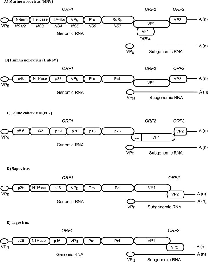
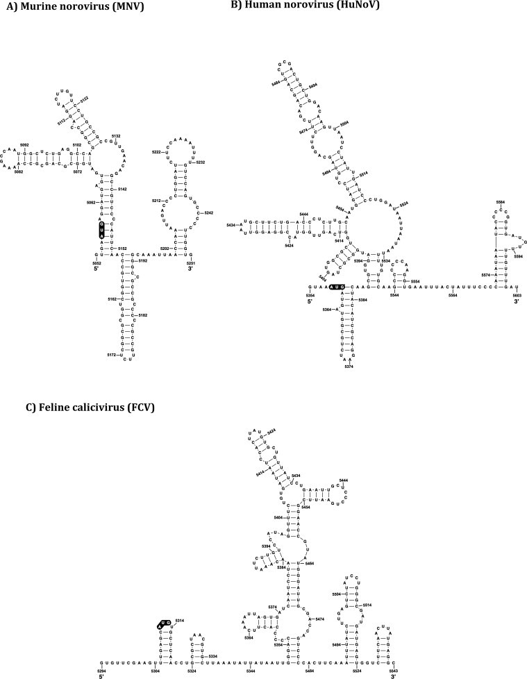
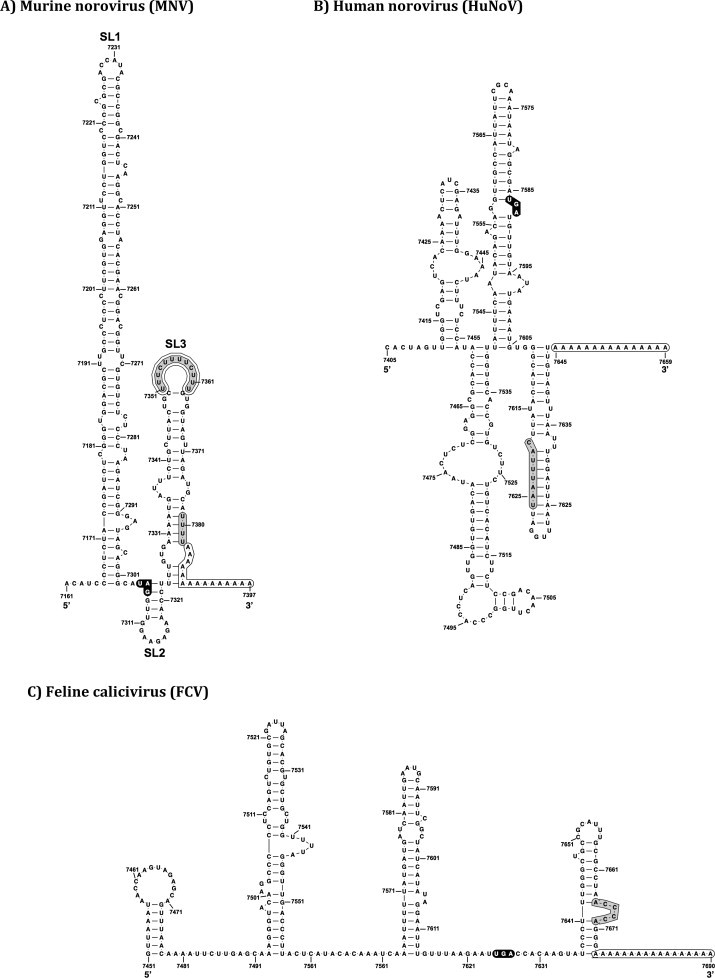
The Caliciviridae family of small positive sense RNA viruses contains a diverse range of pathogens of both man and animals. The molecular mechanisms of calicivirus genome replication and translation have not been as widely studied as many other RNA viruses. With the relatively recent development of robust cell culture and reverse genetics systems for several members of the Caliciviridae family, a more in-depth analysis of the finer detail of the viral life cycle has now been obtained. As a result, the identification and characterization of the role of RNA structures in the calicivirus life cycle has also been possible. This review aims to summarize the current state of knowledge with respect to the role of RNA structures at the termini of calicivirus genomes.

Keywords: Caliciviruses; Norovirus; RNA secondary structures; RNA–protein interactions.


Figures

Fig. 1
Fig. 1
Calicivirus genome organization. The full-length genome is organized into 2–4 ORFs. A subgenomic RNA (sgRNA) consisting of ORF2 and ORF3 is also produced during replication. The viral protein, VPg, is covalently attached to the 5′ end of genomic and sgRNAs and the 3′ end is polyadenylated. ORF1 encodes a polyprotein which is post-translationally cleaved into non-structural proteins, whereas ORF2 and ORF3 encode the structural proteins VP1 and VP2, respectively. The genome layout of (A) MNV, (B) HuNoV, (C) FCV, (D) sapovirus, and (E) lagovirus are shown. The genome of MNV encodes a unique virulence factor (VF1) translated from a recently discovered ORF4. The FCV genome encodes for a leader capsid peptide (LC) at the 5′ end of VP1.
Fig. 2
Fig. 2
Calicivirus 5′ genomic RNA structure. The RNA secondary structures of various caliciviruses, predicted using mfold (Zuker, 2003), and biochemically confirmed for MNV and FCV (Karakasiliotis et al., 2010, Vashist et al., 2012), are shown. The ORF1 initiation codon for each virus is shown in black. The predicted RNA secondary structure are (A) (MNV, GenBank accession number DQ285629), (B) (HuNoVs, GenBank accession number NC_001959), and (C) (FCV, GenBank accession number L40021) 5′ ends. Four potential PTB binding sites (BS1-4) in the FCV genome are highlighted in gray. Note that these are potential binding sites, and only BS2-3 was identified to bind to PTB.
Fig. 3
Fig. 3
Predicted 5′ RNA secondary structures of calicivirus sgRNA. Schematic illustration of the predicted RNA secondary structures of the 5′ sgRNA of (A) MNV, (B) HuNoV, and (C) FCV predicted by mfold (Zuker, 2003). The initiation codon of ORF2 is highlighted in black.
Fig. 4
Fig. 4
RNA structure at the calicivirus 3′ extremity. Schematic representation of the mfold predicted (Zuker, 2003) and biochemically confirmed RNA secondary structures (Bailey et al., 2010) at the 3′ end of calicivirus genomes. The stop codon of the ORF3 is shown in black circles, while the poly A tail is highlighted for each virus at the end of the RNA sequence of the genome. (A) MNV contains three predicted stem-loop structures (SL1-3). The variable p(Y) tract in SL3 is highlighted in gray, and the nucleotides (UUUU) at position 7380 are highlighted in black prior to the poly A tail. In (B) HuNoV and (C) FCV the predicted binding site of the host factor nucleolin is highlighted in gray.

References

    1. Agis-Juárez R.A., Galván I., Medina F., Daikoku T., Padmanabhan R., Ludert J.E., del Angel R.M. Polypyrimidine tract-binding protein is relocated to the cytoplasm and is required during dengue virus infection in Vero cells. J. Gen. Virol. 2009;90:2893–2901. doi: 10.1099/vir.0.013433-0. - DOI - PubMed
    1. Alvarez D.E., Filomatori C.V., Gamarnik A.V. Functional analysis of dengue virus cyclization sequences located at the 5′ and 3′ UTRs. Virology. 2008;375:223–235. doi: 10.1016/j.virol.2008.01.014. - DOI - PubMed
    1. Alvarez D.E., Lodeiro M.F., Ludueña S.J., Pietrasanta L.I., Gamarnik A.V. Long-range RNA–RNA interactions circularize the dengue virus genome. J. Virol. 2005;79:6631–6643. doi: 10.1128/JVI.79.11.6631-6643.2005. - DOI - PMC - PubMed
    1. Bailey D., Karakasiliotis I., Vashist S., Chung L.M.W., Rees J., Reese J., McFadden N., Benson A., Yarovinsky F., Simmonds P., Goodfellow I. Functional analysis of RNA structures present at the 3′ extremity of the murine norovirus genome: the variable polypyrimidine tract plays a role in viral virulence. J. Virol. 2010;84:2859–2870. doi: 10.1128/JVI.02053-09. - DOI - PMC - PubMed
    1. Balinsky C.A., Schmeisser H., Ganesan S., Singh K., Pierson T.C., Zoon K.C. Nucleolin interacts with the dengue virus capsid protein and plays a role in formation of infectious virus particles. J. Virol. 2013;87:13094–13106. doi: 10.1128/JVI.00704-13. - DOI - PMC - PubMed

Publication types

LinkOut - more resources