. 2015 Apr 15;308(8):G691-701.
doi: 10.1152/ajpgi.00116.2014. Epub 2015 Feb 12.

Activation of the renin-angiotensin system stimulates biliary hyperplasia during cholestasis induced by extrahepatic bile duct ligation

Affiliations

Activation of the renin-angiotensin system stimulates biliary hyperplasia during cholestasis induced by extrahepatic bile duct ligation

Syeda H Afroze et al. Am J Physiol Gastrointest Liver Physiol. .

Abstract

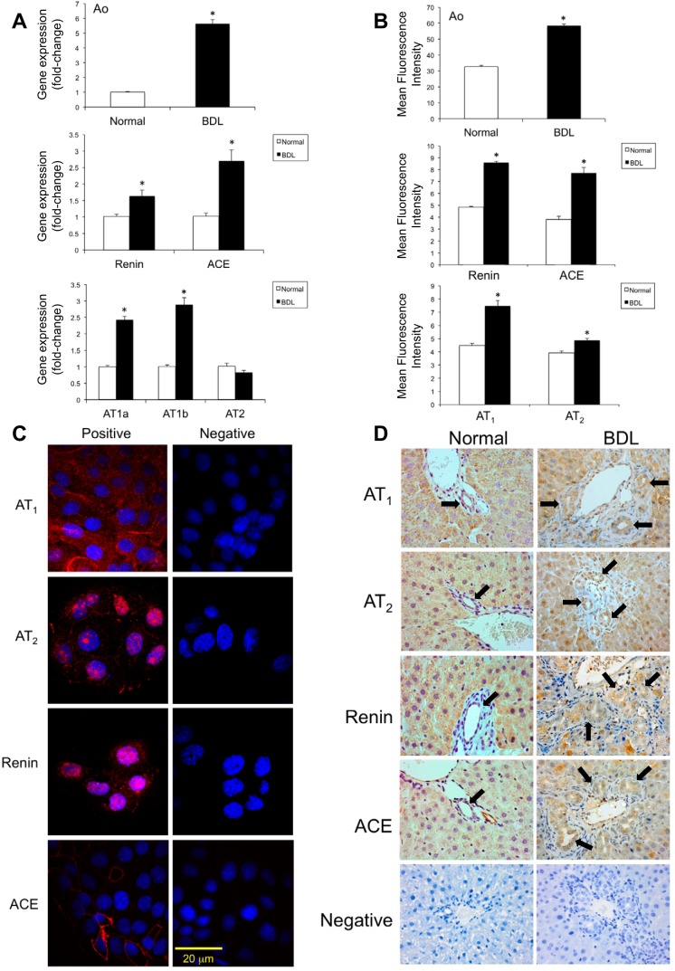
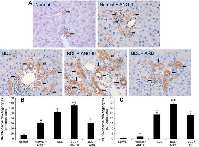
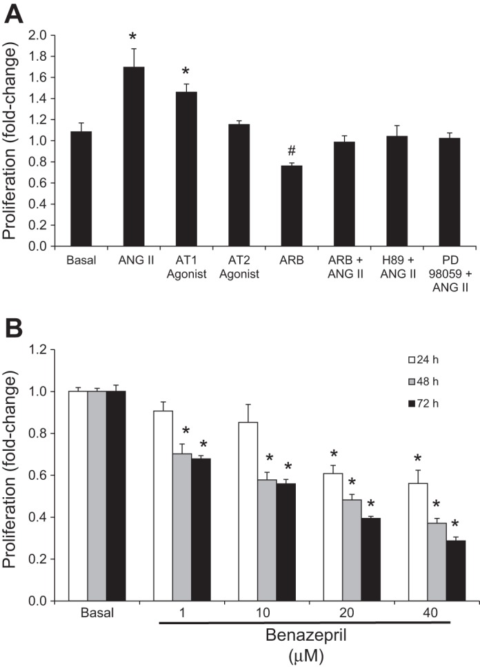
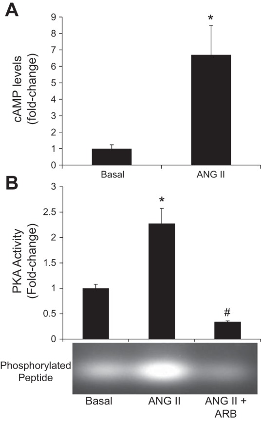
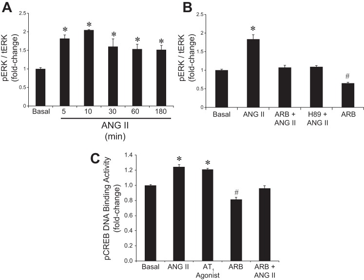
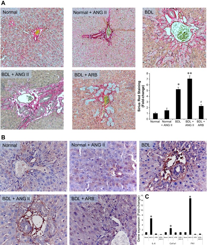
Cholangiocyte proliferation is regulated in a coordinated fashion by many neuroendocrine factors through autocrine and paracrine mechanisms. The renin-angiotensin system (RAS) is known to play a role in the activation of hepatic stellate cells and blocking the RAS attenuates hepatic fibrosis. We investigated the role of the RAS during extrahepatic cholestasis induced by bile duct ligation (BDL). In this study, we used normal and BDL rats that were treated with control, angiotensin II (ANG II), or losartan for 2 wk. In vitro studies were performed in a primary rat cholangiocyte cell line (NRIC). The expression of renin, angiotensin-converting enzyme, angiotensinogen, and angiotensin receptor type 1 was evaluated by immunohistochemistry (IHC), real-time PCR, and FACs and found to be increased in BDL compared with normal rat. The levels of ANG II were evaluated by ELISA and found to be increased in serum and conditioned media of cholangiocytes from BDL compared with normal rats. Treatment with ANG II increased biliary mass and proliferation in both normal and BDL rats. Losartan attenuated BDL-induced biliary proliferation. In vitro, ANG II stimulated NRIC proliferation via increased intracellular cAMP levels and activation of the PKA/ERK/CREB intracellular signaling pathway. ANG II stimulated a significant increase in Sirius red staining and IHC for fibronectin that was blocked by angiotensin receptor blockade. In vitro, ANG II stimulated the gene expression of collagen 1A1, fibronectin 1, and IL-6. These results indicate that cholangiocytes express a local RAS and that ANG II plays an important role in regulating biliary proliferation and fibrosis during extraheptic cholestasis.

Keywords: angiotensin; bile duct ligation; cholangiocyte; renin-angiotensin system.


Figures

Fig. 1.
Fig. 1.
Expression of renin-angiotension system (RAS) components [angiotensinogen (Ao), renin, angiotensin-converting enzyme (ACE), angiotensin receptor types 1a (AT1a), 1b (AT1b), and 2 (AT2)] in liver sections and cholangiocytes isolated from normal and bile duct-ligated (BDL) rats and normal rat intrahepatic cholangiocyte lines (NRIC). RAS components were assessed by fluorescence-activated cell sorting (FACS) analysis (A) and real-time PCR (B) in cholangiocytes from normal and BDL rats. *P < 0.05 vs. normal. Data are presented as means ± SE, n = 6. Expression of RAS components in NRIC cells was evaluated by immunofluorescence (C). Expression of RAS by immunohistochemistry in normal and BDL rat liver sections (D). Original magnification ×40. Black arrows indicate positively stained bile ducts.
Fig. 2.
Fig. 2.
Evaluation of CK-19 and PCNA expression in normal rat (NR) and BDL rat liver sections by immunohistochemistry. A and B: there are increased numbers of CK-19- (A and B) and PCNA-positive (C) cholangioyctes per portal area in Normal + ANG II and BDL + ANG II compared with the NR rat liver. Losartan [angiotensin receptor blocker (ARB)] treatment significantly downregulates the expression of CK-19 and PCNA in isolated cholangiocytes from BDL rats. *P < 0.05 vs. normal. **P < 0.05 vs. BDL. #P < 0.05 vs. BDL. Data are presented as means ± SE, n = 6.
Fig. 3.
Fig. 3.
Evaluation of the effects of angiotensin II (ANG II) on the proliferation of NRICs. A: ANG II and the AT1 agonist stimulate the proliferation of NRIC at 48 h. ANG II-stimulated proliferation is blocked by ARB, H89 (PKA inhibitor), and PD98059 (ERK1/2 pathway inhibitor). ARB (losartan) decreased basal proliferation. *P < 0.05 vs. basal (control). #P < 0.05 vs. basal. Data are presented as means ± SE, n = 6. B: benazepril (ACE inhibitor) dose dependently inhibits NRIC proliferation at 48 and 72 h. *P < 0.05 vs. basal (control). Data are presented as means ± SE, n = 6.
Fig. 4.
Fig. 4.
Effect of ANG II on intracellular cAMP levels and PKA activity in NRICs. A: ANG II stimulated intracellular cAMP levels in NRICs. *P < 0.05 vs. normal. Data are presented as means ± SE, n = 6. B: ANG II stimulated an increase in PKA activity compared with basal, which is prevented by ARB (losartan). *P < 0.05 vs. basal (control). #P < 0.05 vs. basal. Data are presented as means ± SE, n = 6.
Fig. 5.
Fig. 5.
Evaluation of the effects of ANG II on ERK1/2 phosphorylation and pCREB DNA binding activity. A: ANG II significantly induces the phosphorylation of ERK1/2 in NRIC in a time-dependent manner. *P < 0.05 vs. normal. Data are presented as means ± SE, n = 6. B: ANG II-mediated phosphorylation of ERK1/2 is attenuated by pretreatment with ARB (losartan) and H89 (PKA inhibitor) prior to ANG II treatment. ARB alone significantly reduced the phosphorylation of ERK1/2 compared with basal. *P < 0.05 vs. basal (control). #P < 0.05 vs. basal. Data are presented as means ± SE, n = 6. C: ANG II stimulates an increase in DNA binding activity of pCREB, which is blocked by attenuated by ARB (losartan). ARB alone significantly reduced the DNA binding activity of pCREB compared with basal. *P < 0.05 vs. basal (control). #P < 0.05 vs. basal. Data are presented as means ± SE, n = 6.
Fig. 6.
Fig. 6.
Evaluation of Sirius red staining, fibronectin expression in liver sections, and ANG II-induced IL-6 expression in NRIC. A: there is increased levels of fibrosis in the portal areas of the ANG II treated Normal + ANG II and BDL + ANG II compared with the NR rat liver. Losartan (ARB) treatment significantly downregulates fibrosis. Original magnification ×40. B: there is increased fibronectin expression in the portal areas of ANG II treated Normal + ANG II and BDL + ANG II compared with the NR rat liver. Losartan (ARB) treatment downregulates fibronectin expression. Original magnification ×40. C: in vitro, ANG II stimulated the gene expression levels of Col1a1, FN1, and IL-6, which were blocked by ARB. *P < 0.05 vs. normal. **P < 0.05 vs. BDL. #P < 0.05 vs. BDL. Data are presented as means ± SE, n = 6.

References

    1. Agasti AK, Mahajan AU, Phadke AY, Nathani PJ, Sawant P. Comparative randomized study on efficacy of losartan versus propranolol in lowering portal pressure in decompensated chronic liver disease. J Dig Dis 14: 266–271, 2013. - PubMed
    1. Alexander RW, Brock TA, Gimbrone MA Jr, Rittenhouse SE. Angiotensin increases inositol trisphosphate and calcium in vascular smooth muscle. Hypertension 7: 447–451, 1985. - PubMed
    1. Alpini G, Phinizy JL, Glaser S, Francis H, Benedetti A, Marucci L, LeSage G. Development and characterization of secretin-stimulated secretion of cultured rat cholangiocytes. Am J Physiol Gastrointest Liver Physiol 284: G1066–G1073, 2003. - PubMed
    1. Alpini G, Prall R, LaRusso NF. The pathobiology of biliary epithelia. In: The Liver; Biology and Pathobiology, edited by Arias I, Boyer J, Chisari F, Fausto N, Jakoby W, Schachter D, Shafritz DA. Philadelphia, PA: Lippincott Williams & Wilkins, 2001, p. 421–435.
    1. Alvaro D, Mancino MG, Glaser S, Gaudio E, Marzioni M, Francis H, Alpini G. Proliferating cholangiocytes: a neuroendocrine compartment in the diseased liver. Gastroenterology 132: 415–431, 2007. - PubMed

Publication types

MeSH terms

LinkOut - more resources