Review
. 2015 Feb 2:10:975-99.
doi: 10.2147/IJN.S68861. eCollection 2015.

Liposomes as nanomedical devices

Affiliations
Review

Liposomes as nanomedical devices

Giuseppina Bozzuto et al. Int J Nanomedicine. .

Abstract

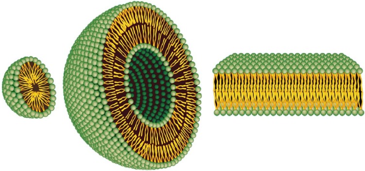
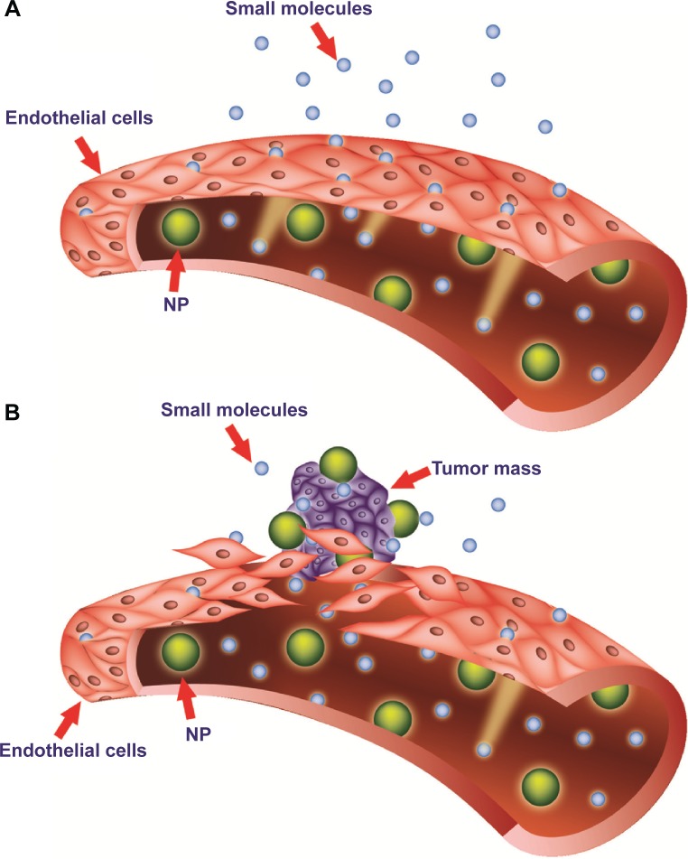
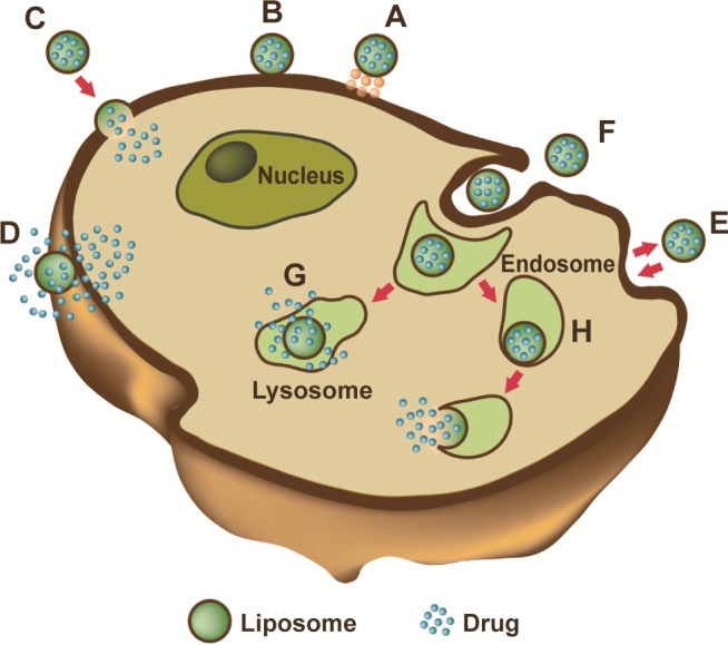
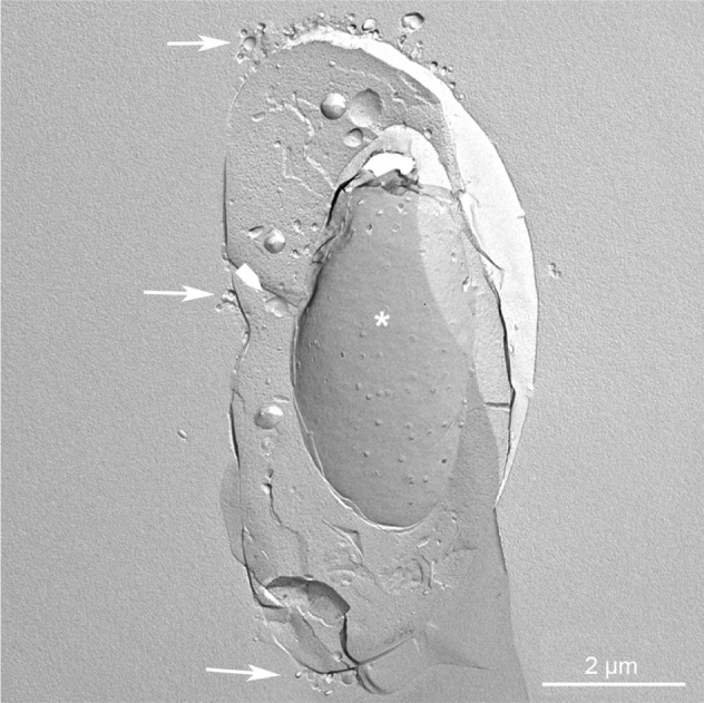
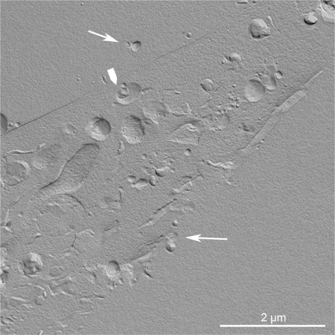
Since their discovery in the 1960s, liposomes have been studied in depth, and they continue to constitute a field of intense research. Liposomes are valued for their biological and technological advantages, and are considered to be the most successful drug-carrier system known to date. Notable progress has been made, and several biomedical applications of liposomes are either in clinical trials, are about to be put on the market, or have already been approved for public use. In this review, we briefly analyze how the efficacy of liposomes depends on the nature of their components and their size, surface charge, and lipidic organization. Moreover, we discuss the influence of the physicochemical properties of liposomes on their interaction with cells, half-life, ability to enter tissues, and final fate in vivo. Finally, we describe some strategies developed to overcome limitations of the "first-generation" liposomes, and liposome-based drugs on the market and in clinical trials.

Keywords: drug delivery; liposomes; nanomedicine; ultrastructure.


Figures

Figure 1
Figure 1
Representation of the steric organization of a micelle (left), a liposome (center), and a lipid bilayer (right). Whereas liposomes are composed of a lipid bilayer, micelles are constructed of one lipid layer in which the apolar section turns inward and the polar heads interact with the environment. These two different organizations mean that the space enclosed in the micelles is much more limited to that available in liposomes. Note: Adapted from Bitounis D, Fanciullino R, Iliadis A, Ciccolini J. Optimizing druggability through liposomal formulations: new approaches to an old concept. ISRN Pharm. 2012;2012:738432.
Figure 2
Figure 2
Targeting of nanomedicines by the enhanced permeability and retention (EPR) effect. Notes: Differences between normal (A) and tumor (B) vessels are depicted. Tumor vessels contain large fenestrations between the endothelial cells: this structural characteristic allows the nanoparticles (NPs) to reach the matrix and the tumor cells by the EPR effect. Conversely, normal tissue contains tightly joined endothelial cells: this prevents the diffusion of NPs outside the blood vessels.
Figure 3
Figure 3
Liposome–cell interaction. Notes: Liposomes loaded with a drug interact with the cell, binding to the surface through receptors (A). Absorption onto the plasma membrane can also occur by electrostatic interactions (B). The delivery of the cargo into the cell cytoplasm can take place through different modes. Lipid nanocarriers fuse with the plasma membrane and discharge drugs into the cell (C). After the interaction with the cell, the structure of the liposome bilayer can be affected and the cargo is released (D). Exchange of carrier-lipid components with the cell membrane can also occur (E). Liposomes internalized by endocytosis (F) can have different fates depending on physicochemical characteristics. Endosomes fuse with lysosomes (G): in this case, the low pH induces the degradation of the liposome membrane and the drug is released. Endosomes follow another route (H): liposomes release their cargo after fusion or the destabilization of the endocytic vesicle.
Figure 4
Figure 4
Replica of a cryofixed and cross-fractured human glioblastoma cell, interacting with dimyristoyl-sn-glycero-phosphatidylcholine liposomes. Liposomes appear mainly clustered on a pole of the cell (arrows). The fracture passes through the cytoplasm, and reveals the nucleus (asterisk) and the cytoplasmic organelles (arrowhead).
Figure 5
Figure 5
Replica of a cryofixed and cross-fractured human glioblastoma cell, interacting with dimyristoyl-sn-glycero-phosphatidylcholine liposomes. Liposomes (arrows) appear in the extracellular space and the cytoplasm of the cell. Some of them are interacting with cytoplasmic organelles (arrowhead).

References

    1. Bangham AD, Horne RW. Negative staining of phospholipids and their structural modification by surface-active agents as observed in the electron microscope. J Mol Biol. 1964;8:660–668. - PubMed
    1. Bangham AD, Hill MW, Miller NG. Preparation and use of liposomes as models of biological membranes. In: Korn ED, editor. Methods in Membrane Biology. Vol. 1. New York: Plenum; 1974. pp. 1–68.
    1. Etheridge ML, Campbell SA, Erdman AG, Haynes CL, Wolf SM, McCullough J. The big picture on nanomedicine: the state of investigational and approved nanomedicine products. Nanomedicine. 2013;9:1–14. - PMC - PubMed
    1. Felice B, Prabhakaran MP, Rodríguez AP, Ramakrishna S. Drug delivery vehicles on a nano-engineering perspective. Mater Sci Eng C Mater Biol Appl. 2014;41:178–195. - PubMed
    1. Fanciullino R, Ciccolini J. Liposome-encapsulated anticancer drugs: still waiting for the magic bullet? Curr Med Chem. 2009;16:4361–4373. - PubMed