Review
. 2014 Dec 2;11(1):26.
doi: 10.1186/2045-8118-11-26. eCollection 2014.

Mechanisms of fluid movement into, through and out of the brain: evaluation of the evidence

Affiliations
Review

Mechanisms of fluid movement into, through and out of the brain: evaluation of the evidence

Stephen B Hladky et al. Fluids Barriers CNS. .

Abstract

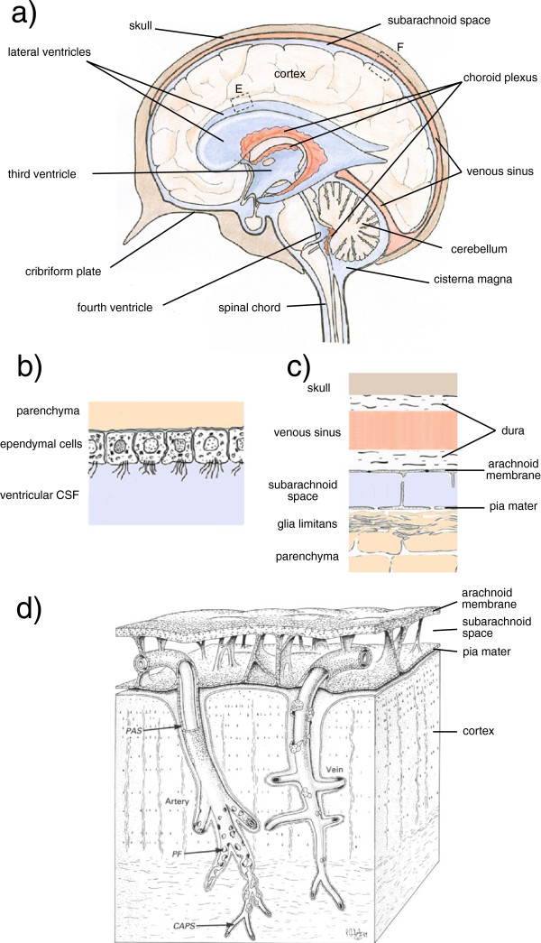
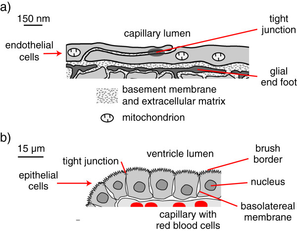
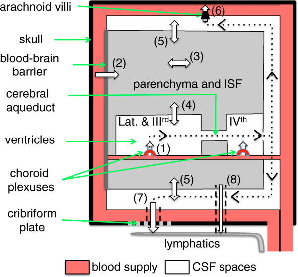
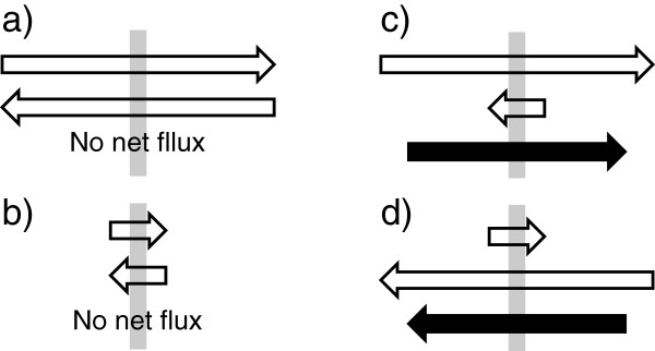
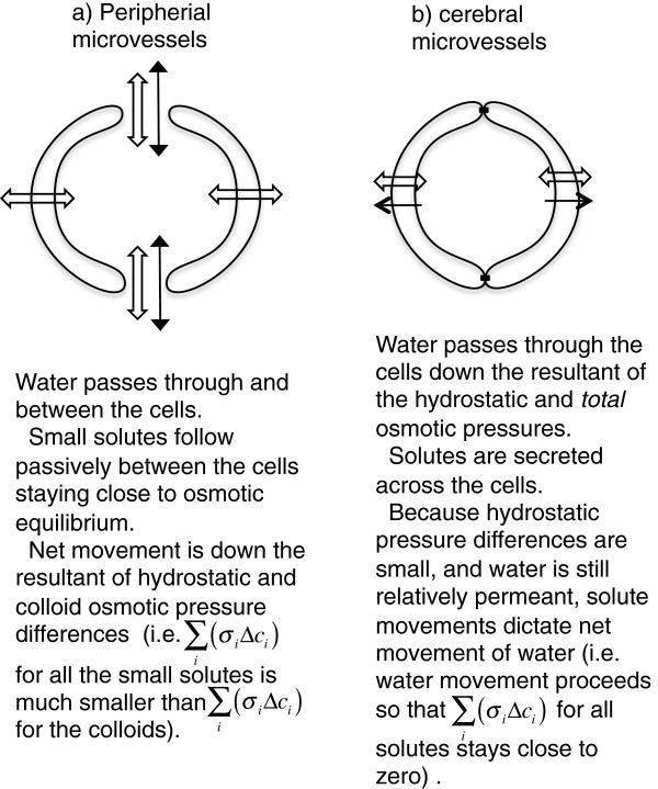
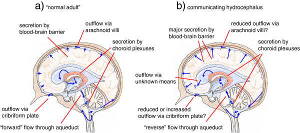
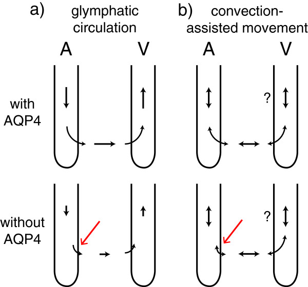
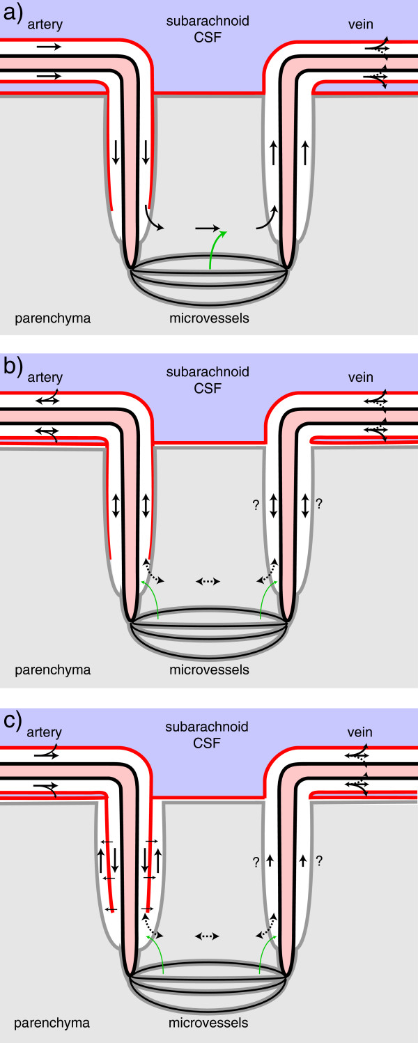
Interstitial fluid (ISF) surrounds the parenchymal cells of the brain and spinal cord while cerebrospinal fluid (CSF) fills the larger spaces within and around the CNS. Regulation of the composition and volume of these fluids is important for effective functioning of brain cells and is achieved by barriers that prevent free exchange between CNS and blood and by mechanisms that secrete fluid of controlled composition into the brain and distribute and reabsorb it. Structures associated with this regular fluid turnover include the choroid plexuses, brain capillaries comprising the blood-brain barrier, arachnoid villi and perineural spaces penetrating the cribriform plate. ISF flow, estimated from rates of removal of markers from the brain, has been thought to reflect rates of fluid secretion across the blood-brain barrier, although this has been questioned because measurements were made under barbiturate anaesthesia possibly affecting secretion and flow and because CSF influx to the parenchyma via perivascular routes may deliver fluid independently of blood-brain barrier secretion. Fluid secretion at the blood-brain barrier is provided by specific transporters that generate solute fluxes so creating osmotic gradients that force water to follow. Any flow due to hydrostatic pressures driving water across the barrier soon ceases unless accompanied by solute transport because water movements modify solute concentrations. CSF is thought to be derived primarily from secretion by the choroid plexuses. Flow rates measured using phase contrast magnetic resonance imaging reveal CSF movements to be more rapid and variable than previously supposed, even implying that under some circumstances net flow through the cerebral aqueduct may be reversed with net flow into the third and lateral ventricles. Such reversed flow requires there to be alternative sites for both generation and removal of CSF. Fluorescent tracer analysis has shown that fluid flow can occur from CSF into parenchyma along periarterial spaces. Whether this represents net fluid flow and whether there is subsequent flow through the interstitium and net flow out of the cortex via perivenous routes, described as glymphatic circulation, remains to be established. Modern techniques have revealed complex fluid movements within the brain. This review provides a critical evaluation of the data.

Keywords: Blood-brain barrier; Brain interstitial fluid; Cerebrospinal fluid; Choroid plexus; Convection; Diffusion; Filtration; Periarterial space; Phase contrast magnetic resonance imaging; Secretion.


Figures

Figure 1
Figure 1
Structures of the brain considered in this review. a) Mid-sagittal section from nose to the back of the head incorporating images of the lateral ventricles that lie to each side of the section. CSF filled spaces are shown in blue, blood filled spaces in pale red. The choroid plexuses are shown in darker red. b) Enlarged view of surface of a lateral ventricle at (E). c) Enlarged view of the cortical surface at (F). The glia limitans is a mat of glial processes. d) Drawing showing the relationships between the arachnoid membrane, the pia mater, the subarachnoid space, and the vasculature supplying the cortex. As described by Zhang et al. for the artery “the sheath has been cut away to show that the periarterial spaces (PAS) of the intracerebral and extracerebral arteries are in continuity. The layer of pial cells becomes perforated (PF) and incomplete as smooth muscle cells are lost from the smaller branches of the artery. The pial sheath finally disappears as the perivascular spaces are obliterated around capillaries (CAPS). Perivascular spaces around the vein (right of picture) are confluent with the subpial space and only small numbers of pial cells are associated with the vessel wall” [23]. The cortical boundary along these vessels is formed by glial foot-processes. The graphic elements in a) and b) and in c) with minor extensions are taken with permission from Figure one of Strazielle et al.[19] and relabelled. d) is reproduced with permission from Figure ten in [23] and partially relabelled. R.O. Weller (personal communication) has emphasized that the spaces shown between the arterial wall and the pial sheath and between the sheath and the glial end-feet were virtual spaces in their electron micrographs. The periarterial spaces are also portrayed in Figure 6.
Figure 2
Figure 2
Key features of a) blood-brain barrier and b) choroid plexus. a) Sketch based on electron micrographs [34] of brain microvessel walls showing endothelial cells linked by tight junctions. These cells form a complete lining of the microvessel lumen and are in turn surrounded by glial end-feet. Occasional pericytes embedded in the basement membrane that separates the endothelial cells and the end-feet are not shown. The tight junctions limit the transport of solutes between the endothelial cells but the basement membrane, the gaps between the glial end-feet and the extracellular spaces within the parenchyma allow passage of molecules as big as proteins. Mitochondria occupy up to 10 % of the endothelial cell volume [35] and provide a substantial source of energy for transport. b) Diagram of one side of a choroid plexus villus as seen in light micrographs (e.g. [2]). Note the 100-fold difference in scales. Each choroid plexus has many such villi, each consisting of a layer of epithelial cells linked by tight junctions and surrounding a core of connective tissue containing capillaries. The endothelial cell layer lining these capillaries is not sealed by tight junctions. The epithelial cells have a prominent apical brush border facing the ventricle and a band of tight junctions separating the apical and basolateral domains of the cell membrane. There is folding of the basolateral membranes, which like the individual microvilli of the brush border are not visible in light micrographs (for electron micrographs see Figures two & twelve in [15]). The microvilli and basolateral folds increase the surface area of the membrane domains and hence the number of transporters that can be located on the two sides of the cells. Choroid plexus epithelium has the structure and properties of a leaky epithelium capable of transporting large quantities of isosmotic fluid.
Figure 3
Figure 3
Schematic plan of the whole brain indicating the fluid movements considered in this review. Fluid is secreted into the ventricles across the choroid plexuses (1) and into the brain parenchyma across the blood-brain barrier (2). Fluid components can move through the parenchyma (3) and there are exchanges of water and solutes (4) and (5) between the interstitial fluid (ISF) of the parenchyma and cerebrospinal fluid (CSF) contained in the ventricles and in the subarachnoid spaces respectively. There is net fluid outflow across the arachnoid villi (6) leading to the dural venous sinuses (including but not restricted to the superior sagittal sinus) and along cranial nerves, most notably the olfactory nerve leading to the cribriform plate (7) and thence to the nasal mucosa. There may also be outflow of fluid in the walls of arteries or veins (8) leading to lymph nodes in the neck. The traditional view of the directions of net CSF flow is indicated by the dotted lines with arrowheads.
Figure 4
Figure 4
Fluxes inwards or outwards across a membrane compared with net flux. The membranes are indicated by the grey bars. In a) and b) the magnitudes of the fluxes are very different but in both cases the fluxes in the two directions are equal and hence there is no net flux. In c) and d) the magnitudes of the fluxes in the two directions are very different and there is a net flux in the direction indicated by the black arrow. Although the flux from left to right is the same in a) and c), because the fluxes in the opposite direction are not the same there is a net flux in c) but not in a). Hence net flux cannot be inferred from measurements of flux in a single direction.
Figure 5
Figure 5
Comparisons of fluid transport across peripheral and cerebral microvessels. In the periphery a) small solutes cross the vessel walls via gaps between the cells. Small solute movement is rapid, therefore their concentration gradients are small and thus unlike the large solutes, the colloids, the small solutes do not oppose the movements of water. Thus except during brief transients, e.g. when osmotic gradients are artificially imposed, net fluid movement is governed by the hydrostatic and colloid osmotic pressure differences between blood and surrounding tissues. In the brain b) paracellular movement is limited by tight junctions. Thus small solutes cross the vessel walls only slowly and the direction and extent of their movement is determined by specific transporters. Hence they are as effective as the large solutes in producing osmotic gradients that dictate the extent of water movement into or out of the surrounding tissue.
Figure 6
Figure 6
Diagram indicating positions within the cortical parenchyma of periarterial spaces that may allow fluid movement. The spaces shown correspond to those around the arteries in Figure 1d. The diagram has been formatted so that it may be compared with earlier published versions [1]. The diagram (not to scale) shows: the tunica intima, the endothelial lining of the lumen and a covering of elastic tissue; the tunica media, a smooth muscle layer; and the tunica adventitia, mainly connective tissue. Within these layers there are two possible free spaces. The inner, called the periarterial space by Zhang et al.[23] and labelled the inner periarterial space in the diagram, is continuous with the periarterial space of the subarachnoid portion of the same artery. The outer that in contact with the brain parenchyma, is likely to be the space described by His [1]. In the view of Weller and associates (personal communication) both the inner periarterial space and the space of His are virtual spaces with no thickness and fluid movement occurs preferentially in the extracellular spaces of the smooth muscle layer. Other interpretations of the spaces are considered in section 4.3.4.
Figure 7
Figure 7
View of net CSF flow pattern in normal adult brain compared with that proposed for communicating hydrocephalus. a) In the normal adult, most of the CSF is secreted by the choroid-plexuses and is reabsorbed through the arachnoid villi or via the cribriform plate leading to the nasal mucosa. There is modest secretion across the blood-brain barrier into ISF most of which emerges into CSF across brain surfaces. The net flow through the cerebral aqueduct is from the third ventricle towards the fourth ventricle. b) In communicating hydrocephalus, the observation of reverse net flow through the aqueduct implies that formation of CSF occurs outside of the ventricles, probably by more extensive fluid secretion across the blood-brain barrier into the cortex and thence out into the CSF-containing spaces. Reverse net flow also requires some route for removal of CSF from the third and lateral ventricles. This route must accommodate both fluid secreted by the choroid plexuses located in these ventricles and fluid entering the third ventricle via the aqueduct. Possible routes are discussed in section 4.2.4. (The background image is the same as in Figure one-a taken from Strazielle et al. with permission [19]).
Figure 8
Figure 8
Effect of absence of AQP4 on fluid flow along perivascular routes. In both a) the glymphatic circulation proposal and b) the convection-assisted movement hypotheses described in Figure 9, the absence of AQP4 results in reduced movement of fluid from the periarterial spaces into the parenchyma (red arrow). In the glymphatic circulation proposal this would produce a build-up of fluid in the periarterial space, which would reduce flow into that space and would also remove the source of fluid destined for perivenous outflow. If instead, as in the convection-assisted movement hypotheses, fluid flow can occur in both directions in the periarterial spaces with little net flow, absence of AQP4 may not affect fluid flow along the periarterial space, a result more consistent with the data. A indicates the periarterial spaces, V the perivenous spaces. The question marks indicate that convection-assisted movement may or may not also occur in the perivenous spaces.
Figure 9
Figure 9
Possible schemes to explain rapid influx of markers via periarterial spaces: a) the glymphatic proposal based on Figure five of Iliff et al. [[143]], b) stirring or mixing and c) layered flow. In a) and c) there is preferential influx via the space between the arterial wall and the pial sheath (the inner periarterial space in Figure 6) while in b) convection back and forth, speeds up the rate of transfer of markers in both directions. Red lines represent pial membranes, grey lines the layer of glial end-feet or glia limitans, solid arrows are fluxes of markers carried or assisted by convection, dashed arrows are either by diffusion or assisted by convection and green arrows have been added as a reminder that fluid secreted by the blood-brain barrier contributes to the fluid in the parenchyma. The location of the pial barriers is based on Zhang et al.[23] (Figures 1d and 6). In c) the influx of fluid via the periarterial spaces may inflate the space of His providing a route for the return of fluid to the subpial space at the cortical surface and then to CSF in the subarachnoid space.

References

    1. Woollam DHM, Millen JW. Perivascular spaces of the mammalian central nervous system. Biol Rev Camb Philos Soc. 1954;29:251–283. doi: 10.1111/j.1469-185X.1954.tb00597.x. - DOI
    1. Millen J, Woollam D. The Anatomy of the Cerebrospinal Fluid. London: Oxford University Press; 1962.
    1. Hayman LA, Weathers SW, Kirkpatrick JB. Atlas of cerebrospinal fluid spaces. In: Hayman LA, Hinck VC, editors. Clinical Brain Imaging: Normal Structure and Functional Anatomy. St. Louis: Mosby-Year Book; 1992. pp. 306–328.
    1. Cserr HF. Physiology of choroid plexus. Physiol Rev. 1971;51:273–311. - PubMed
    1. Welch K. The principles of physiology of the cerebrospinal fluid in relation to hydrocephalus including normal pressure hydrocephalus. Adv Neurol. 1975;13:247–332. - PubMed