

Comparing the monoisocentric and dual isocentric techniques in chest wall radiotherapy of mastectomy patients
- PMID: 25679164
- PMCID: PMC5689976
- DOI: 10.1120/jacmp.v16i1.5069
Comparing the monoisocentric and dual isocentric techniques in chest wall radiotherapy of mastectomy patients
Abstract
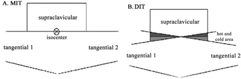
The monoisocentric (MIT) and dual isocentric (DIT) techniques are compared for the mastectomy patients undergoing chest wall radiotherapy, and a new practical method is suggested for determining the dose calculation reference point to be used in the MIT. Data of 18 mastectomy patients having chest wall radiotherapy were used. To find the appropriate dose calculation reference point for the MIT, the target tissue was divided into nine regions with 17 points as the appropriate candidates. After finding the best reference point for the MIT, dose calculations were made for each patient based on the MIT and DIT to determine the dose distributions of the target volume and organs at risk. The lateral component of the dose calculation reference point was found to be located at one-third of the distance between the geometrical center and the lateral border of the chest wall in the lateral direction toward the outer border. The longitudinal component of this point was found to be located at the geometrical center of the chest wall with a depth located around 2-3 cm under the patients' skin. There was no significant difference between the two radiotherapy planning techniques (MIT and DIT) regarding the dose distributions in the organs at risk and the 95% of the prescribed dose coverage of the target tissue. However, a significant difference for the 105% of the prescribed dose coverage, maximum dose delivered to the target tissue, and the level 2 lymph nodes dose was found, with the DIT showing higher values. Because of the good matching and no superposition observed between the treatment fields in the MIT, it was expected and confirmed that the hot and cold regions (with higher and lower doses than the prescribed dose) with the MIT are significantly fewer than that of the DIT. Therefore, to perform a better conformal radiotherapy for the patients having mastectomy, it could be recommended to use the MIT instead of the DIT and other conventional techniques.
Figures


References
-
- Halperin EC, Perez CA, Brady LW, Wazer DE, Freeman C, Prosnitz LR. Perez and Brady's principles and practice of radiation oncology, 5th ed. Philadelphia, PA: Lippincott Williams & Wilkins; 2007.
-
- Khan FM. Treatment planning in radiation oncology, 2nd ed. Philadelphia, PA: Lippincott Williams & Wilkins; 2007.
-
- Marshall MG. Three‐field isocentric breast irradiation using asymmetric jaws and a tilt board. Radiother Oncol. 1993;28(3):228–32. - PubMed
-
- Edlund T and Gannett D. A single isocentric technique using CT‐based planning in the treatment of breast cancer. Med Dosim. 1999;24(4):239–45. - PubMed
-
- Hernandez V, Arenas M, Pons F, Sempau J. Clinical applications of geometrical field matching in radiotherapy based on a new analytical solution. Med Dosim. 2011;36(2):160–65. - PubMed
Publication types
MeSH terms
LinkOut - more resources
Full Text Sources
Other Literature Sources
Medical
