

Multiple hepatic sclerosing hemangioma mimicking metastatic liver tumor successfully treated by laparoscopic surgery: Report of a case
- PMID: 25679307
- PMCID: PMC4353964
- DOI: 10.1016/j.ijscr.2015.01.032
Multiple hepatic sclerosing hemangioma mimicking metastatic liver tumor successfully treated by laparoscopic surgery: Report of a case
Abstract
Introduction: Hepatic sclerosing hemangioma is a very rare benign tumor, characterized by fibrosis and hyalinization occurring in association with degeneration of a hepatic cavernous hemangioma. We report here a rare case of multiple hepatic sclerosing hemangioma mimicking metastatic liver tumor that was successfully treated using laparoscopic surgery.
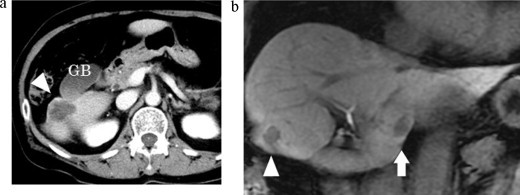
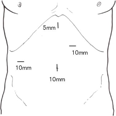
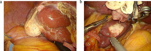
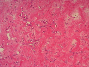
Presentation of case: A 67-year-old woman with multiple liver tumors underwent single-incision laparoscopic sigmoidectomy under a diagnosis of advanced sigmoid cancer with multiple liver metastases. Examination of surgical specimens of sigmoid colon revealed moderately differentiated adenocarcinoma invading the serosa, and no lymph node metastases. Serum levels of carcinoembryonic antigen and carbohydrate antigen 19-9 remained within normal limits throughout the course. Two months after sigmoidectomy, the patient underwent laparoscopic partial hepatectomy of S1 and S6 of the liver and cholecystectomy. Histopathological examination showed that the tumors mainly comprised hyalinized tissue and collagen fibers with sporadic vascular spaces on hematoxylin and eosin-stained sections, yielding a diagnosis of multiple hepatic sclerosing hemangioma. No evidence of recurrence has been seen as of 21 months postoperatively.
Discussion: Differentiating multiple sclerosing hemangiomas from metastatic liver tumors was quite difficult because the radiological findings were closely compatible with liver metastases. Laroscopic hepatectomy provided less blood loss, a shorter duration of hospitalization, and good cosmetic results.
Conclusion: Sclerosing hemangioma should be included among the differential diagnoses of multiple liver tumors in patients with colorectal cancer. Laparoscopic hepatectomy is useful for diagnostic therapy for undiagnosed multiple liver tumors.
Keywords: Hepatic sclerosing hemangioma; Laparoscopic liver resection; Metastatic liver tumor.
Copyright © 2015 The Authors. Published by Elsevier Ltd.. All rights reserved.
Figures
References
-
- Iwahashi S., Shimada M., Utsunomiya T., Imura S., Morine Y., Ikemoto T. Laparoscopic hepatic resection for metastatic liver tumor of colorectal cancer: comparative analysis of short- and long-term results. Surg. Endosc. 2014;28:80–84. - PubMed
-
- Takayasu K., Moriyama N., Shima Y., Muramatsu Y., Yamada T., Makuuchi M. Atypical radiographic findings in hepatic cavernous hemangioma: correlation with histologic features. AJR Am. J. Roentgenol. 1986;146:1149–1153. - PubMed
-
- Haratake J., Horie A., Nagafuchi Y. Hyalinized hemangioma of the liver. Am. J. Gastroenterol. 1992;87:234–236. - PubMed
-
- Cheng H.C., Tsai S.H., Chiang J.H., Chang C.Y. Hyalinized liver hemangioma mimicking malignant tumor at MR imaging. AJR Am. J. Roentgenol. 1995;165:1016–1017. - PubMed
LinkOut - more resources
Full Text Sources
Other Literature Sources
Research Materials
