. 2015 Mar;156(3):439-450.
doi: 10.1097/01.j.pain.0000460325.25762.c0.

Modulation of nociceptive dural input to the trigeminocervical complex through GluK1 kainate receptors

Affiliations

Modulation of nociceptive dural input to the trigeminocervical complex through GluK1 kainate receptors

Anna P Andreou et al. Pain. 2015 Mar.

Abstract

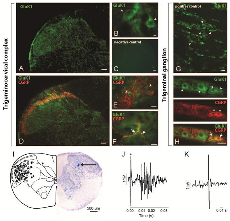
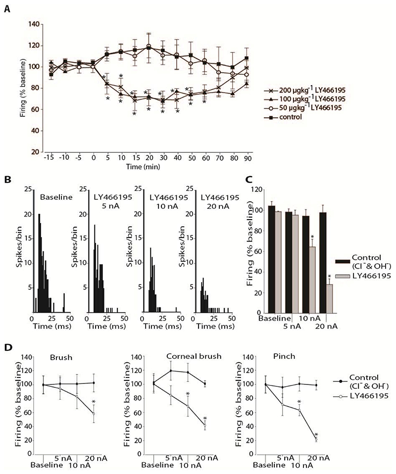
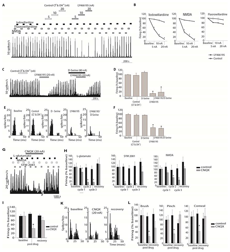
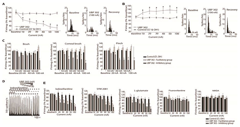
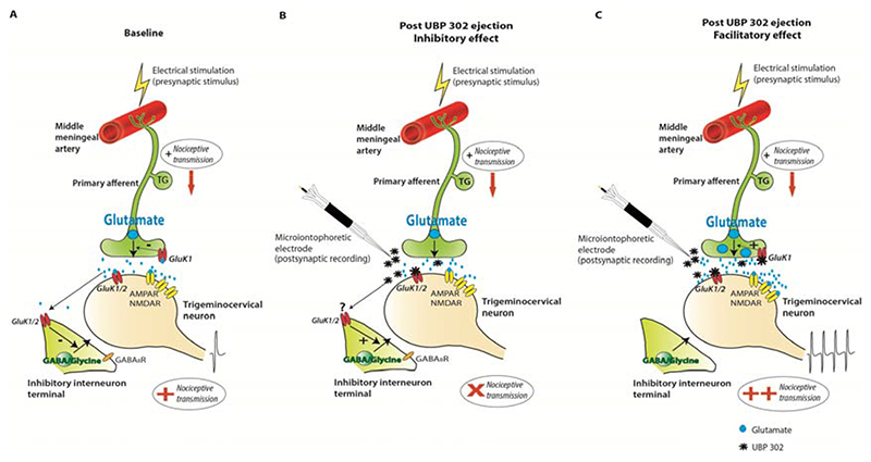
Migraine is a common and disabling neurologic disorder, with important psychiatric comorbidities. Its pathophysiology involves activation of neurons in the trigeminocervical complex (TCC). Kainate receptors carrying the glutamate receptor subunit 5 (GluK1) are present in key brain areas involved in migraine pathophysiology. To study the influence of kainate receptors on trigeminovascular neurotransmission, we determined the presence of GluK1 receptors within the trigeminal ganglion and TCC with immunohistochemistry. We performed in vivo electrophysiologic recordings from TCC neurons and investigated whether local or systemic application of GluK1 receptor antagonists modulated trigeminovascular transmission. Microiontophoretic application of a selective GluK1 receptor antagonist, but not of a nonspecific ionotropic glutamate receptor antagonist, markedly attenuated cell firing in a subpopulation of neurons activated in response to dural stimulation, consistent with selective inhibition of postsynaptic GluK1 receptor-evoked firing seen in all recorded neurons. In contrast, trigeminovascular activation was significantly facilitated in a different neuronal population. The clinically active kainate receptor antagonist LY466195 attenuated trigeminovascular activation in all neurons. In addition, LY466195 demonstrated an N-methyl-d-aspartate receptor-mediated effect. This study demonstrates a differential role of GluK1 receptors in the TCC, antagonism of which can inhibit trigeminovascular activation through postsynaptic mechanisms. Furthermore, the data suggest a novel, possibly presynaptic, modulatory role of trigeminocervical kainate receptors in vivo. Differential activation of kainate receptors suggests unique roles for this receptor in pro- and antinociceptive mechanisms in migraine pathophysiology.


Conflict of interest statement

Conflicts of interest: None

The other authors declare no competing financial interests.

Figures

Figure 1
Figure 1. GluK1-like immunofluorescent staining within the trigeminocervical complex and trigeminal ganglia, and reconstruction of recording sites.
A-B. Photomicrographs of the trigeminocervical complex taken from the C1level, showing GluK1-like staining (green). Cell bodies and punctate staining was evident in laminae I-III and mainly cell bodies were seen in deeper laminae. Stained cells were round or pear-shaped. D-F. Within lamina I and outer lamina II, where calcitonin gene-related peptide (CGRP)-like fibers project (red), GluK1-like staining (green) was seen in cell bodies as well as in punctate staining, which might represent proximal processes or GluK1 primary fibers as some co-localization (yellow) with CGRP was seen (Figure 1G). C. A photomicrograph of a negative control section of the TCC. G. A photomicrograph of GluK1-like cells (green) within the trigeminal ganglion. H. GluK1-like cells (green) mainly co-localized with small size CGRP-positive cells (red) within the trigeminal ganglion. Arrowheads present examples of GluK1-like positive cells, stars indicate CGRP-like positive cells and arrows show co-localization of CGRP- and GluK1-like fibers (F) or cells (H). Scale bars = 100 μm (A, C, D), 50 μm (E, G, H), 10 μm (B, F). I. Reconstruction of recording sites within the C1 spinal cord level, plotted after Paxinos and Watson [43], identified histologically (solid circles represent pontamine sky blue spots) and by microdrive readings (open circles). A photomicrograph demonstrating an original recording site marked by ejection of pontamine sky blue (arrow) is shown. J. An original trace showing a cluster of cells in the trigeminocervical complex, firing in response to stimulation of the middle meningeal artery (100 μs, 0.5 Hz, 12 volts; * stimulus artefact). K. Original tracing from a neuron in the trigeminocervical complex responding to microiontophoresis of L-glutamate.
Figure 2
Figure 2
A. Effect of intravenously administrated LY466195 on responses of trigeminocervical neurons to middle meningeal artery stimulation. LY466195 inhibited firing to dural electrical stimulation at 200 and 100 μgkg-1, but not at 50 μgkg-1. Saline control ejection had no effect on neuronal firing. * P < 0.05 B. Effect of microiontophoresis of LY466195 on responses of trigeminocervical neurons to middle meningeal artery stimulation and to receptive field characterisation. Example of post-stimulus histograms from a representative neuron, recorded during baseline conditions and during ejection of LY466195 at currents 5, 10 and 20 nA. C. Comparison of the middle meningeal artery stimulation evoked firing under each condition. Ejection of LY466195 demonstrated a dose dependent inhibition of the cell firing, maximally at 20 nA. D. Effects of microiontophoretically delivered LY466195 and its vehicle control (Cl-, OH-) on the firing rates of second order neurons in response to receptive field characterisation. Microiontophoretic application of LY466195 at 20 nA significantly inhibited firing to both innocuous (brush) and nocuous (corneal brush; pinch) stimulation of the ophthalmic dermatome, while a lower ejection current (10 nA) significantly inhibited noxious stimulation of the receptive field. * P < 0.05
Figure 3
Figure 3
A. Example of the effects of LY466195 on the firing rates to pulsed ejections of Fluorowillardiine, Iodowillardiine and NMDA. Cell firing in response to the ejected agonists returned to baseline levels within 2-5 minutes after LY466195 microiontophoresis ceased. B. Comparison of the effect of LY466195 and control ions (Cl-, OH-) on iodowillardiine, NMDA and fluorowillardiine evoked responses. Cell firing in response to iodowillardiine and NMDA was significantly inhibited by microiontophoretically administered LY466195. C. Example of the response of a trigeminocervical neuron to pulsed NMDA. Ejection of serine, LY466195 and control ions are shown with the solid bars. Ejection of LY466195 demonstrated a potent inhibitory effect on NMDA-evoked firing that was not reversed by pre-treatment with serine. D. Comparison of the NMDA evoked firing during ejection of control ions and LY466195. E. Post-stimulus histograms generated from a representative trigeminocervical neuron following electrical stimulation of the middle meningeal artery. Microiontophoresis of serine or control ions at the same current had no effect on neuronal firing. Co-ejection of serine with LY466195 failed to block the inhibitory actions of LY466195. F. Comparison of the middle meningeal artery stimulation-evoked firing under the influence of LY466195, serine and control ions. G. Example of the effects of CNQX on the firing rates to pulsed ejections of the L-glutamate, Iodowillardiine and SYM 2081. Cell firing in response to the ejected agonists returned to baseline levels within 30 min after CNQX microiontophoresis ceased. B-H. Effects of CNQX on cell firing in response to L-glutamate, SYM 2081 and NMDA, separately for each ejection cycle. CNQX strongly inhibited L-glutamate, SYM 2081 and NMDA-evoked firing. Ejection of control ions had no significant effects on glutamate agonists-evoked firing. I. Comparison of the middle meningeal artery stimulation-evoked firing under the influence of CNQX and control ions. CNQX significantly inhibited neuronal firing in response to trigeminovascular stimulation. K. Post-stimulus histograms generated from a representative trigeminocervical neuron following electrical stimulation of the middle meningeal artery during baseline conditions, microiontophoresis of CNQX and recovery. neuronal firing. L. Effects of microiontophoretically delivered CNQX on the firing rates of second order neurons in response to receptive field characterisation. Microiontophoretic application of CNQX at 20 nA significantly inhibited firing to both innocuous (brush) and nocuous (corneal brush; pinch) stimulation of the ophthalmic dermatome, while a lower ejection current (10 nA) significantly inhibited noxious stimulation of the receptive field. * P < 0.05 compared to control
Figure 4
Figure 4
Effect of microiontophoresis of UBP302 on responses of trigeminocervical neurons to middle meningeal artery stimulation and to receptive field characterisation. Ejection of UBP302 produced two opposing responses. A. In 15 units ejection of UBP302 demonstrated a dose dependent inhibition of the cell firing, maximally at 100 nA. B. In 12 neurons UBP302 significantly potentiated cell firing. Control vehicle had no effect. Post-stimulus histograms from representative cluster of neurons in the inhibitory (A) and facilitatory (B) groups, recorded pre- UBP302 ejection, during ejection of UBP302 at 100 nA, and 30 minutes after the cessation of ejection. C. Effects of microiontophoretically delivered UBP302 and its vehicle control (Cl-, OH-) on the firing rates of second order neurons in response to receptive field characterisation. Cell firing in response to light brush was not altered by microiontophoretically administered UBP302 neither in the inhibitory group nor in the facilitatory group. Cell firing in response to pinch and to corneal stimulation was significantly inhibited by microiontophoretically administered UBP302 in the inhibitory group. Noxious-evoked responses to both pinch and corneal stimulus were not significantly facilitated across the cohort in the facilitatory group. Control ejection had no significant effect. D. Example of the effects of UBP302 on the firing rates to pulsed ejections of fluorowillardiine, iodowillardiine and NMDA. Cell firing in response to the ejected agonists returned to baseline levels within 30 minutes after UBP302 microiontophoresis ceased. E. Comparison of the current-response curves for UBP302 and control (Cl- and OH-) on iodowillardiine, SYM 2081, L-glutamate , fluorowillardiine and NMDA evoked responses. Overall, cell firing in response to iodowillardiine (n = 19), SYM 2081 (n = 18) and L-glutamate (n = 18) was significantly inhibited by microiontophoretically administered UBP302, and there was no significant difference between the inhibitory and the facilitatory groups. Cell firing in response to fluorowillardiine (n = 17) and NMDA (n = 6) was unaffected by microiontophoretically delivered UBP302. Vehicle control had no significance on any agonist-evoked firing. * P < 0.05
Figure 5
Figure 5. Proposed mechanism of action of microiontophoresed UBP302 on trigeminovascular activation.
Electrical stimulation of the middle meningeal artery will activate fibers arising from the trigeminal ganglion (TG) and increase neuronal firing of second order neurons in the trigeminocervical complex. A. During baseline recordings, activation of primary afferents innervating the middle meningeal artery causes glutamate release in the trigeminocervical complex. Glutamate activates post-synaptic kainate receptors in addition to AMPA and NMDA receptors activation, promoting trigeminocervical nociceptive transmission. Pre-synaptic kainate receptors on primary trigeminal afferents could be activated by glutamate and control glutamate release, whereas activation of kainate receptors on inhibitory interneurons is also possible, and will modulate inhibitory synaptic transmission. B. Local application of UBP302 by microiontophoresis can selectively block post-synaptic kainate receptors on second order neurons and inhibit nociceptive transmission, most likely in the absence of kainate receptors on the pre-synaptic trigeminal nerve fiber. Blockade of kainate receptors on inhibitory GABAergic terminals could occur due to UBP302 diffusion, and this could additionally account for the resultant analgesic effect. However, as we did not directly study this, such a blockade of kainate heteroreceptors on GABAergic interneurons is shown with a “?”. In such a synaptic environment post-synaptic kainate receptors play an important role in trigeminovascular nociceptive transmission and their selective blockade could prove a beneficial treatment for migraine. C. In a different synaptic environment where both pre- and post-synaptic kainate receptors are present on trigeminal fibers and second-order trigeminocervical neurons respectively, local application of UBP302 by microiontophoresis can selectively block post-synaptic kainate receptors on trigeminocervical neurons. In addition blockade of pre-synaptic kainate receptors on primary afferents might inhibit the negative control feedback of glutamate release by kainate receptors. This results in increased glutamate release upon stimulation of the trigeminal fibers innervating the middle meningeal artery. Released glutamate could then act on a bigger scale on AMPA and NMDA receptors and facilitate nociceptive transmission. In these neurons it is also possible that post-synaptic GluK1 kainate receptors do not mediate sensory transmission.

References

    1. Afridi S, Giffin NJ, Kaube H, Goadsby PJ. A randomized controlled trial of intranasal ketamine in migraine with prolonged aura. Neurology. 2013;80:642–647. - PubMed
    1. Agrawal SG, Evans RH. The primary afferent depolarizing action of kainate in the rat. Br J Pharmacol. 1986;87(2):345–355. - PMC - PubMed
    1. Akerman S, Goadsby PJ. Topiramate inhibits cortical spreading depression in rat and cat: impact in migraine aura. Neuroreport. 2005;16(12):1383–1387. - PubMed
    1. Akerman S, Goadsby PJ. Topiramate inhibits trigeminovascular activation: an intravital microscopy study. Br J Pharmacol. 2005;146(1):7–14. - PMC - PubMed
    1. Andreou AP, Goadsby PJ. Therapeutic potential of novel glutamate receptor antagonists in migraine. Expert opinion on investigational drugs. 2009;18(6):789–803. - PubMed

Publication types

MeSH terms

Substances