

Cone beam computed tomography assessment of the maxillary incisive canal and foramen: considerations of anatomical variations when placing immediate implants
- PMID: 25679505
- PMCID: PMC4332502
- DOI: 10.1371/journal.pone.0117251
Cone beam computed tomography assessment of the maxillary incisive canal and foramen: considerations of anatomical variations when placing immediate implants
Abstract
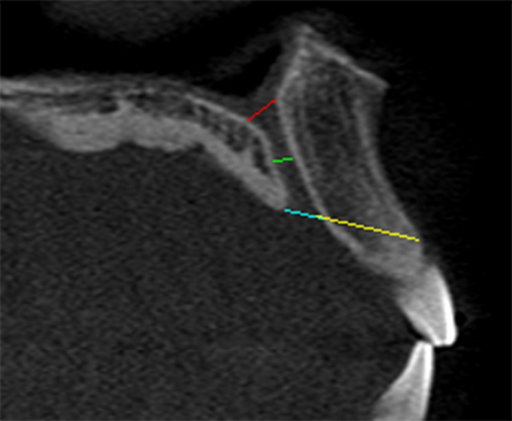
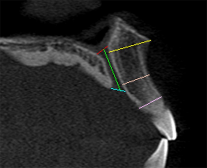
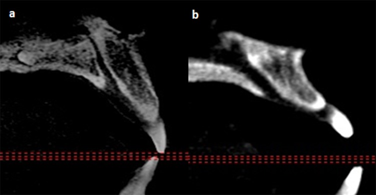
Introduction: The maxillary incisive canal connects the roof of the oral cavity with the floor of nasal cavity and has the incisive and nasal foramina respectively at its two opposite ends. Its close proximity with the anterior incisors affects one's ability to place immediate implants in ideal position.
Objective: To avoid causing complication, variations in their dimensions were studied.
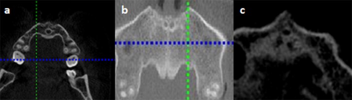
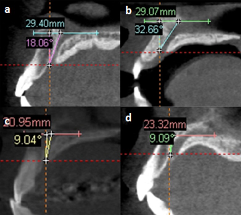
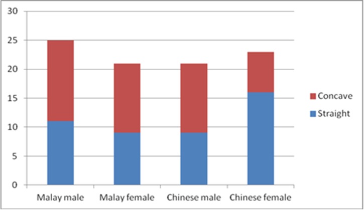
Material and methods: Images of ninety Mongoloids patients examined with i-CAT Cone Beam Computed Tomography were included. The sizes of the nasopalatine foramen, the incisive canal and foramen, and anterior maxillary bone thickness were measured. The direction and course of the canals were assessed.
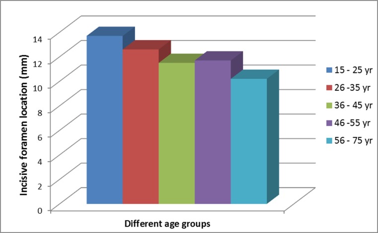
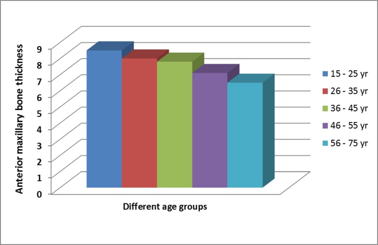
Results: The mean labiopalatal and mesiodistal measurements of the incisive foramen were 2.80 mm and 3.49 mm respectively, while the labiopalatal width of the nasal foramen was 6.06 mm. The incisive canal was 16.33 mm long and 3.85 mm wide. The anterior maxillary bone has an average thickness of 7.63 mm. The dimensions of the incisive foramen and incisive canal, and anterior maxillary bone thickness demonstrated gender differences with males showing greater values. The anterior maxillary bone thickness was affected by age but this difference was not observed in canal dimensions. The majority of subjects have a funnel shape-like incisive canal with the broader opening located at its superior. They seem to have a longer slanted-curve canal with one channel at its middle portion and a narrower incisive foramen opening than those reported elsewhere.
Conclusions: This study found that gender is an important factor that affected the characteristics of the IC and the amount of bone anterior to it. Male generally had bigger IC and thicker anterior bone. In addition, the anterior maxillary bone thickness was affected by aging, where it becomes thinner with increased age even though the subjects were fully dentate.
Conflict of interest statement
Figures
References
-
- Sarandha DL, Hussain ZU (2008) Textbook of Complete Denture Prosthodontics. Jaypee Brothers, Medical Publishers;
-
- Scheid RC, Woelfel JB (2007) Woelfel's Dental Anatomy: Its Relevance to Dentistry. Lippincott Williams & Wilkins; 10.1093/jxb/erm028 - DOI
-
- Drake R, Vogl AW, Mitchell AWM (2009) Gray's Anatomy for Students. Elsevier Health Sciences;
-
- Ghom AG (2008) Normal Radiographic Anatomy. In: Ghom A.G. eds., Textbook of Oral Radiology. New Delhi: Elsevier: pp. 191–212.
-
- Glickman GN (2002) Preparation for Treatment. In: Cohen S., Burns R. C. eds., Pathway of the Pulp. St Louis: Mosby: pp. 77–109.
Publication types
MeSH terms
LinkOut - more resources
Full Text Sources
Other Literature Sources
Miscellaneous
