

Human vagus nerve branching in the cervical region
- PMID: 25679804
- PMCID: PMC4332499
- DOI: 10.1371/journal.pone.0118006
Human vagus nerve branching in the cervical region
Abstract
Background: Vagus nerve stimulation is increasingly applied to treat epilepsy, psychiatric conditions and potentially chronic heart failure. After implanting vagus nerve electrodes to the cervical vagus nerve, side effects such as voice alterations and dyspnea or missing therapeutic effects are observed at different frequencies. Cervical vagus nerve branching might partly be responsible for these effects. However, vagus nerve branching has not yet been described in the context of vagus nerve stimulation.
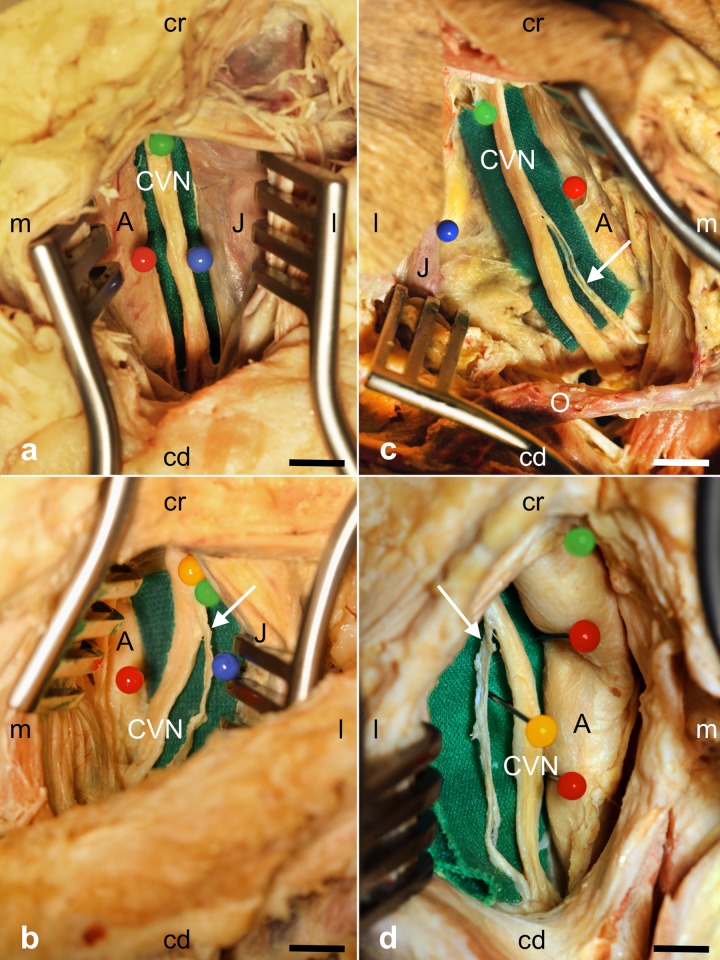
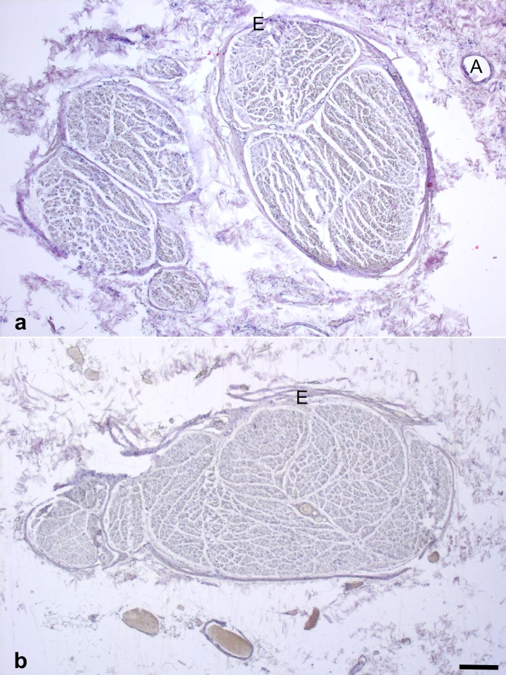
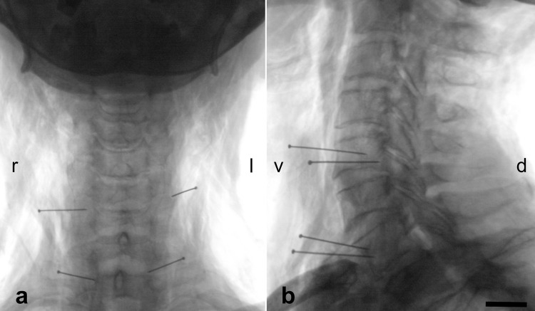
Materials and methods: Branching of the cervical vagus nerve was investigated macroscopically in 35 body donors (66 cervical sides) in the carotid sheath. After X-ray imaging for determining the vertebral levels of cervical vagus nerve branching, samples were removed to confirm histologically the nerve and to calculate cervical vagus nerve diameters and cross-sections.
Results: Cervical vagus nerve branching was observed in 29% of all cases (26% unilaterally, 3% bilaterally) and proven histologically in all cases. Right-sided branching (22%) was more common than left-sided branching (12%) and occurred on the level of the fourth and fifth vertebra on the left and on the level of the second to fifth vertebra on the right side. Vagus nerves without branching were significantly larger than vagus nerves with branches, concerning their diameters (4.79 mm vs. 3.78 mm) and cross-sections (7.24 mm2 vs. 5.28 mm2).
Discussion: Cervical vagus nerve branching is considerably more frequent than described previously. The side-dependent differences of vagus nerve branching may be linked to the asymmetric effects of the vagus nerve. Cervical vagus nerve branching should be taken into account when identifying main trunk of the vagus nerve for implanting electrodes to minimize potential side effects or lacking therapeutic benefits of vagus nerve stimulation.
Conflict of interest statement
Figures
References
-
- Alexopoulos AV, Kotagal P, Loddenkemper T, Hammel J, Bingaman WE (2006) Long-term results with vagus nerve stimulation in children with pharmacoresistant epilepsy. Seizure 15: 491–503. - PubMed
-
- Coady MA, Adler F, Davila JJ, Gahtan V (2000) Nonrecurrent laryngeal nerve during carotid artery surgery: case report and literature review. J Vasc Surg 32: 192–196. - PubMed
MeSH terms
LinkOut - more resources
Full Text Sources
Other Literature Sources
