. 2015 Feb 13;10(2):e0117158.
doi: 10.1371/journal.pone.0117158. eCollection 2015.

Two novel mutations in myosin binding protein C slow causing distal arthrogryposis type 2 in two large Han Chinese families may suggest important functional role of immunoglobulin domain C2

Affiliations

Two novel mutations in myosin binding protein C slow causing distal arthrogryposis type 2 in two large Han Chinese families may suggest important functional role of immunoglobulin domain C2

Xuefu Li et al. PLoS One. .

Erratum in

Abstract

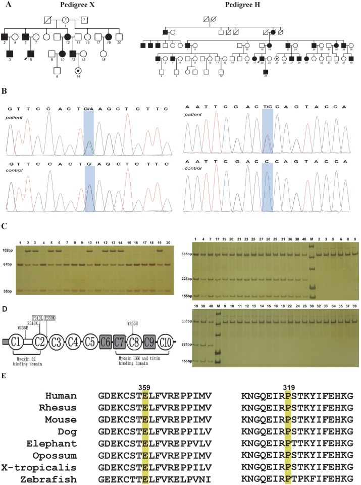
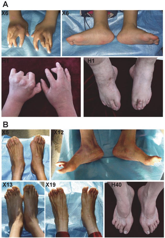
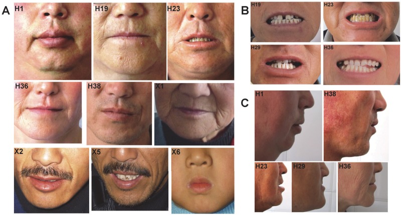
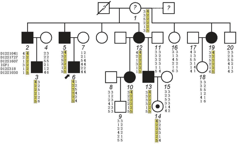
Distal arthrogryposes (DAs) are a group of disorders that mainly involve the distal parts of the limbs and at least ten different DAs have been described to date. DAs are mostly described as autosomal dominant disorders with variable expressivity and incomplete penetrance, but recently autosomal recessive pattern was reported in distal arthrogryposis type 5D. Mutations in the contractile genes are found in about 50% of all DA patients. Of these genes, mutations in the gene encoding myosin binding protein C slow MYBPC1 were recently identified in two families with distal arthrogryposis type 1B. Here, we described two large Chinese families with autosomal dominant distal arthrogryposis type 2(DA2) with incomplete penetrance and variable expressivity. Some unique overextension contractures of the lower limbs and some distinctive facial features were present in our DA2 pedigrees. We performed follow-up DNA sequencing after linkage mapping and first identified two novel MYBPC1 mutations (c.1075G>A [p.E359K] and c.956C>T [p.P319L]) responsible for these Chinese DA2 families of which one introduced by germline mosacism. Each mutation was found to cosegregate with the DA2 phenotype in each family but not in population controls. Both substitutions occur within C2 immunoglobulin domain, which together with C1 and the M motif constitute the binding site for the S2 subfragment of myosin. Our results expand the phenotypic spectrum of MYBPC1-related arthrogryposis multiplex congenita (AMC). We also proposed the possible molecular mechanisms that may underlie the pathogenesis of DA2 myopathy associated with these two substitutions in MYBPC1.


Conflict of interest statement

Competing Interests: The authors have declared that no competing interests exist.

Figures

Fig 1
Fig 1. MYBPC1 mutations in the DA2 families.
A: Chinese kindreds affected with DA2. Solid and open symbols represent affected and unaffected individuals, respectively. The small filled circle inside the open circle represents an asymptomatic carrier. The question mark indicates the unknown status of the individuals of family X. The numbers denote individuals whose DNA samples were available for the analysis. Dotted line in pedigree X indicates individuals 16, 19 and 20 were born from different fathers. B: The c. 1075G>A (p.E359K) mutation in pedigree X and c. 956C>T (p.P319L) mutation in pedigree H of MYBPC1. Sanger sequence analysis of an affected individual and a normal unaffected control. The mutations shown as a light blue shadow each. C: Digests of MYBPC1 amplicons from family members. In pedigree X, the mutation eliminates an Ecl136II restriction site. Digests of MYBPC1 amplicons (102 bp) from affected individuals fractionate into three fragments (102 bp, 67 bp and 35 bp), whereas only two fragments (67 bp and 35 bp) were found in the unaffected members and the mildly symptomatic founder (X1). In pedigree H, the mutation creates a novel Hinf I restriction site. Digests of MYBPC1 amplicons (421 bp) from affected individuals fractionate into four fragments (383 bp, 228 bp, 155 bp, and 38 bp), whereas the 38-bp product cannot be observed on this gel. Only one fragment (383 bp) is observed in unaffected individuals. M indicates DNA marker. D: Schematic demonstrating the position of the mutations in the MYBPC1 protein. Two DA2 MYBPC1 mutations reported in the present study, p.P319L and p.E359K are shown in rectangles. Similar to the mutation of R318X found in patients with lethal congenital contracture syndrome type 4, both of them are located within the C2 immunoglobulin domain. The immunoglobulin domains C1, C2 and the MyBP-C unique motif (M-motif) between them constitute a critical region, which has been shown to interact with myosin S2. The DA1 MYBPC1 mutations p.W236R and p.Y856H, located within the M-motif and the C8 repeat respectively, are also shown. Immunoglobulin domains are shown as open circles and fibronectin type III domains as grey squares. M-motif between C1 and C2 domain is shown as open rectangle. E: Amino acid alignment around the each affected residue of the MYBPC1 protein. The highly conserved E359 and P319 are highlighted in yellow.
Fig 2
Fig 2. Typical limb contractures and overextension contractures in the two DA2 families.
A: Note that typical limb contractures include ulnar deviation of fingers, adducted stiff/clasped thumbs, severe camptodactyly, overlapping fingers (H1), vertical talus(X6) and clubfeet after surgical corrections accompanied by overriding toes (H1) in severely affected individuals. B: Note that overextension contractures were observed at the metatarsophalangeal joints(X5, X12 and H40) or the proximal interphalangeal joints(X13 and X19) of the toes.
Fig 3
Fig 3. Facial contractures in affected individuals from both Chinese DA2 families.
A: Note that prominent nasolabial folds and small nose with small nostrils were present in the affected individuals. Pouting lips (H1, H23, X2, X5 and X6) or pinched lips (H19, H36 and X1) were noticed. A crease extending laterally and downward from corners of the mouth (prominent in H1 and X1, mild in H19, H23 and H36) or “non-H-shaped” cutaneous dimples on the sides of the chin (H38, X2 and X5) were also observed. The prominent nasolabial folds extended downward below the corners of the mouth so that which exhibiting like “parentheses” around the mouth (X2 and X5). A very small whistling mouth (X6) and a deep vertical groove besides the left corner of the mouth (H38) were remarkable. B: Note that dental crowding were found in four affected individuals of pedigree H. C: Micrognathia was also present in pedigree H.
Fig 4
Fig 4. Haplotype analysis in pedigree X.
Haplotype analysis indicating microsatellite markers on chromosome 12q flanking MYBPC1 common to all affected individuals with distal arthrogryposis type 2. Common haplotype is highlighted in yellow and derived from the mildly symptomatic founder (case X1). The question mark indicates the unknown status of the individuals of family X. Case X1 had minor facial anomalies, suggesting she may have a somatic mosaicism of the MYBPC1 mutation (p.E359K). The small filled circle inside the open circle indicates an asymptomatic carrier. Marker’s name is shown at the left. Dotted line in pedigree X indicates individuals 16, 19 and 20 were born from different fathers.

Similar articles

Cited by

References

    1. Bamshad M, Jorde LB, Carey JC (1996) A revised and extended classification of the distal arthrogryposes. Am J Med Genet 65: 277–281. - PubMed
    1. Hall JG, Reed SD, Greene G (1982) The distal arthrogryposes: delineation of new entities—review and nosologic discussion. Am J Med Genet 11: 185–239. - PubMed
    1. Dieterich K, Quijano-Roy S, Monnier N, Zhou J, Faure J, et al. (2013) The neuronal endopeptidase ECEL1 is associated with a distinct form of recessive distal arthrogryposis. Hum Mol Genet 22: 1483–1492. 10.1093/hmg/dds514 - DOI - PubMed
    1. Beck AE, McMillin MJ, Gildersleeve HI, Kezele PR, Shively KM, et al. (2013) Spectrum of mutations that cause distal arthrogryposis types 1 and 2B. Am J Med Genet A 161A: 550–555. 10.1002/ajmg.a.35809 - DOI - PMC - PubMed
    1. Stevenson DA, Carey JC, Palumbos J, Rutherford A, Dolcourt J, et al. (2006) Clinical characteristics and natural history of Freeman-Sheldon syndrome. Pediatrics 117: 754–762. - PubMed

Publication types

MeSH terms

Supplementary concepts

LinkOut - more resources