Review
. 2015 Feb:32:113-20.
doi: 10.1016/j.ceb.2015.01.004. Epub 2015 Feb 11.

Desmin related disease: a matter of cell survival failure

Affiliations
Review

Desmin related disease: a matter of cell survival failure

Yassemi Capetanaki et al. Curr Opin Cell Biol. 2015 Feb.

Abstract

Maintenance of the highly organized striated muscle tissue requires a cell-wide dynamic network that through interactions with all vital cell structures, provides an effective mechanochemical integrator of morphology and function, absolutely necessary for intra-cellular and intercellular coordination of all muscle functions. A good candidate for such a system is the desmin intermediate filament cytoskeletal network. Human desmin mutations and post-translational modifications cause disturbance of this network, thus leading to loss of function of both desmin and its binding partners, as well as potential toxic effects of the formed aggregates. Both loss of normal function and gain of toxic function are linked to mitochondrial defects, cardiomyocyte death, muscle degeneration and development of skeletal myopathy and cardiomyopathy.


Figures

Figure 1
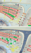
Figure 1. Schematic representation of the desmin intermediate filament scaffold with its direct and indirect interactome in cardiac muscle (A) and the consequences due to its deficiency or disruption in desmin related disease (B)
A. Desmin, the muscle-specific IF (in yellow) together with its associated non-muscle specific IFs (synemin, syncoilin, paranemin in blue) form a continuous network linking the contractile apparatus at the Z-disc level with different membranous compartments (intercalated discs and costameres) and organelles (nucleus, mitochondria; interaction with other organelles, e.g. sarcoplasmic reticulum (SR) and lysosomes are not shown). Most of the proteins mediating the direct association of desmin with the above compartments respectively (desmoplakin, myospryn, ankyrin, plectin) are shown. Desmin associates with the chaperon αB-crystallin in multiple compartments. Several other desmin-associated proteins (e.g. hsp27, nebulin etc.) are not shown. B. The catastrophic effects of desmin deficiency or disruption of desmin network due to its mutations, PTMs and TNF-α induced cleavage [11••] are depicted. Desmin aggregates, the hallmark of DRMs, often containing high amounts of many other proteins (see text) (here only αB-crystallin and filamin C) are shown. Fragmented and degenerating mitochondria is the first and most prominent defect; disruption of the association of desmin IFs with the nucleus (nuclear deformation), defects of the ID and mislocalization of its components to lateral sarcolemma regions are shown. Myospryn looses completely its perinuclear localization in the absence of desmin [56•]. Increased ECM and fibrosis due to inflammation (not shown) triggered by cardiomyocyte death is evident between cells.
Figure 2
Figure 2. A. Structural organization of the human desmin and the localization of identified disease causing mutations throughout its molecule
The desmin molecule, like all IFs, has a long highly conserved central α-helical rod domain, interrupted by 3 short nonhelical linkers (L1, L12, L2), thus generating 4 helices (1A, 1B, 2A, 2B), responsible for coiled-coil dimer formation. The central rod is flanked by nonhelical head (amino-terminal) and tail (carboxy-terminal) domains that are sites of post-translational modifications. Numbers indicate the amino-acid position of the domain borders. 68 pathogenic desmin mutations have been reported so far. The majority resides within the 2B helical domain and the tail. Most of them (53 out of total 68) are missense mutations, leading to single aminoacid substitutions but some are small in-frame deletions (e.g. Glu359_Ser361del) and frame-shift mutations (e.g. Lys241GlufsX4). Mutations with cardiac manifestation are indicated with different colored dots. DCM causing mutations are shown with blue, HCM with green, RCM with orange and ARVD/C with purple. Mutations that affect known phosphorylation sites (Ser7Phe and Ser13Phe) are indicated with solid red (P), while mutations on potential phosphorylation sites with dotted red (P). Two known substrates of ADP-Ribosylation (Arg58 and Arg73) at the head domain are indicated with green (R). TNFα-induced caspase 6 cleavage of desmin at Met263 within the L12 linker domain [11••] is shown; the Ile451Met mutation at the tail domain that promotes the proteolytic cleavage of desmin at the N-terminus [19] is indicated with asterisc (*). B. Diagram summarizing the mechanisms causing desmin network deregulation and the cellular defects due the loss of function of desmin and desmin-associated proteins, eventually causing desmin related pathology.
Figure 3
Figure 3. TNF-α-induced heart failure is the most common DRM
TNF-α-induced caspase cleavage of desmin leads to aggregate formation (asterisk) and loss of its intercalated disc (ID) localization, as shown by immunofluorescence analysis of desmin (green) on representative TNF-α (right panel) and WT (left panel) myocardial sections. Arrow: Z-lines, arrowhead: IDs. Nuclei are stained with DAPI (blue). Adopted from [11]. Scalebar: 20 μm.

Similar articles

Cited by

References

    1. Ho CY, et al. Lamin A/C and emerin regulate MKL1-SRF activity by modulating actin dynamics. Nature. 2013;497(7450):507–11. This paper describes for the first time the involvement of the nuclear intermediate filament proteins, lamins, in the modulation of the mechanochemical transcription factor MKL1. - PMC - PubMed
    1. Swift J, et al. Nuclear lamin-A scales with tissue stiffness and enhances matrix-directed differentiation. Science. 2013;341(6149):1240104. The importance of nuclear lamins in a systematic relationship between tissue mechanics, retinoic acid nuclear entry and stem cell differentiation was described for the first time. - PMC - PubMed
    1. Capetanaki Y, et al. Muscle intermediate filaments and their links to membranes and membranous organelles. Exp Cell Res. 2007;313(10):2063–76. - PubMed
    1. Capetanaki Y. Desmin cytoskeleton: a potential regulator of muscle mitochondrial behavior and function. Trends Cardiovasc Med. 2002;12(8):339–48. - PubMed
    1. Selcen D. Myofibrillar myopathies. Neuromuscul Disord. 2011;21(3):161–71. - PMC - PubMed

Publication types