. 2015 Feb 13;10(2):e0118203.
doi: 10.1371/journal.pone.0118203. eCollection 2015.

Consistent inhibition of cyclooxygenase drives macrophages towards the inflammatory phenotype

Affiliations

Consistent inhibition of cyclooxygenase drives macrophages towards the inflammatory phenotype

Yi Rang Na et al. PLoS One. .

Abstract

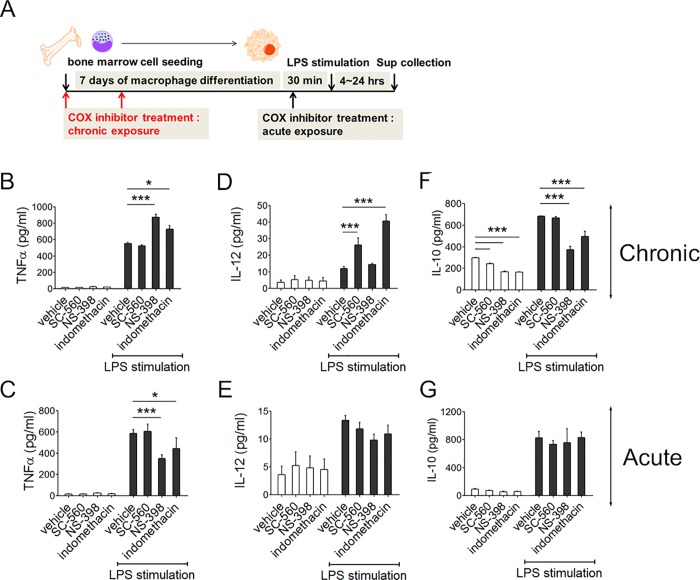
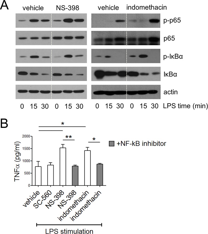
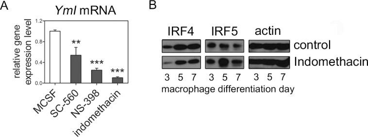
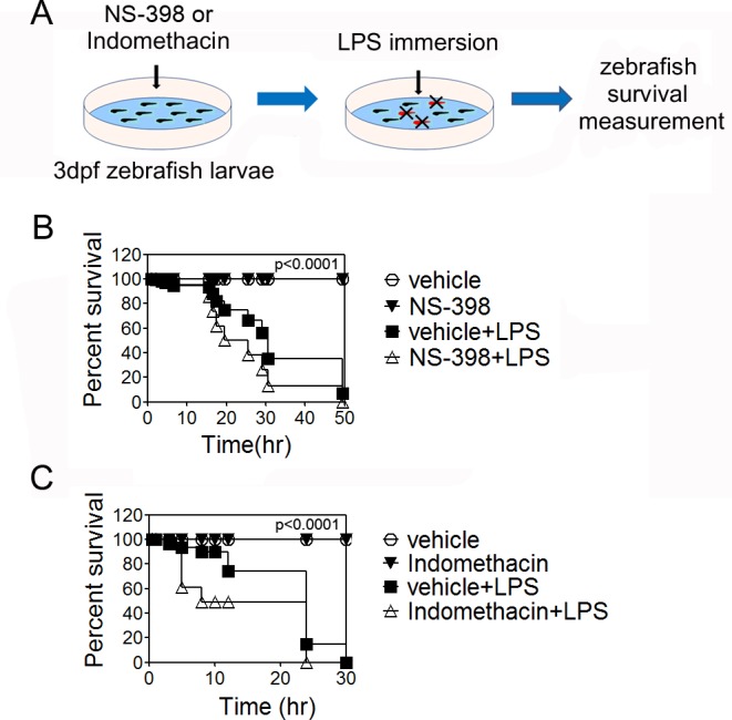
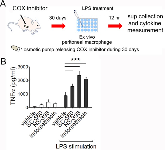
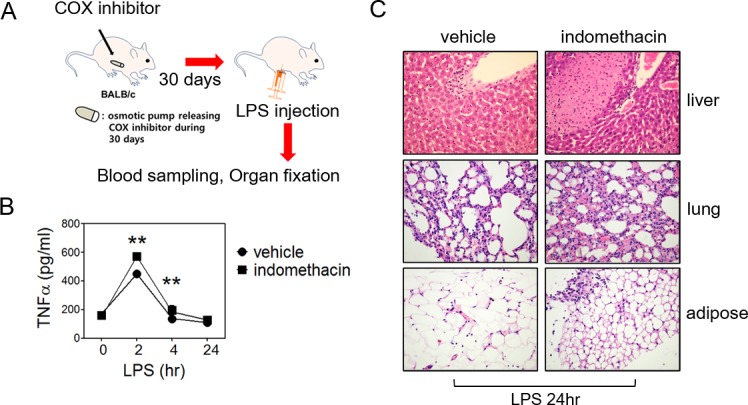
Macrophages play important roles in defense against infection, as well as in homeostasis maintenance. Thus alterations of macrophage function can have unexpected pathological results. Cyclooxygenase (COX) inhibitors are widely used to relieve pain, but the effects of long-term usage on macrophage function remain to be elucidated. Using bone marrow-derived macrophage culture and long-term COX inhibitor treatments in BALB/c mice and zebrafish, we showed that chronic COX inhibition drives macrophages into an inflammatory state. Macrophages differentiated in the presence of SC-560 (COX-1 inhibitor), NS-398 (COX-2 inhibitor) or indomethacin (COX-1/2 inhibitor) for 7 days produced more TNFα or IL-12p70 with enhanced p65/IκB phosphoylation. YmI and IRF4 expression was reduced significantly, indicative of a more inflammatory phenotype. We further observed that indomethacin or NS-398 delivery accelerated zebrafish death rates during LPS induced sepsis. When COX inhibitors were released over 30 days from an osmotic pump implant in mice, macrophages from peritoneal cavities and adipose tissue produced more TNFα in both the basal state and under LPS stimulation. Consequently, indomethacin-exposed mice showed accelerated systemic inflammation after LPS injection. Our findings suggest that macrophages exhibit a more inflammatory phenotype when COX activities are chronically inhibited.


Conflict of interest statement

Competing Interests: The authors have declared that no competing interests exist.

Figures

Fig 1
Fig 1. Cytokine responses in COX inhibited bone-marrow-derived macrophages.
Panel A: Time line of macrophage differentiation with COX inhibition and LPS stimulation. COX inhibitors were treated in two modes: SC-560 (COX-1 inhibitor), NS-398 (COX-2 inhibitor) or indomethacin (COX-1/2 inhibitor) were treated at 30 min before LPS stimulation (acute) or during whole differentiation period (chronic). Panels B-G: Macrophage TNFα (Panels B, C), IL-12p70 (Panels D, E) and IL-10 (Panels F, G) productions analyzed by ELISA. COX inhibitors were treated chronically (Panels B, D, F) or acutely (Panels C, E, G). LPS was treated at 100 ng/ml and culture medium was collected after 4 h (TNFα) and 24 h (IL-12p70 and IL-10) incubation for analysis. Results represent means ± SE of three independent experiments. Statistical analysis was performed by one-way ANOVA. *P<0.05, ***P<0.001.
Fig 2
Fig 2. Analysis of TLR4 early signaling pathways in COX inhibited macrophages.
Panel A: Phospho-IκBα, total IκBα, phospho-p65, total p65 and actin protein levels from whole cell lysates were detected by Western blotting. Macrophages were differentiated for 7 days in the presence of NS-398 or indomethacin and compared with the control at the indicated time points after LPS stimulation. Each compared lanes for one protein all come from same blots. Panel B: NF-κB inhibitor (20 ng/ml) was treated to chronically NS-398 or indomethacin treated macrophages 30 min before LPS stimulation. LPS was treated at 100 ng/ml and culture medium was collected after 4 h incubation for analysis. Results represent means ± SE of three experiments. Statistical analysis was performed by one-way ANOVA. *P<0.05, **P<0.005.
Fig 3
Fig 3. YmI, IRF4 and IRF5 expressions in COX-inhibited murine macrophages.
Bone-marrow-derived macrophages were differentiated in the presence of indicative COX inhibitors for 7 days. Panel A: Real-time PCR was performed to examine YmI expression. Results represent relative fold changes compared with the control after normalization to endogenous GAPDH expression. Three independent experiments were performed. **P<0.01, ***P<0.001 Panel B: Western blotting representing IRF4, IRF5 and actin in macrophages at differentiation day 3, 5 and 7 with or without indomethacin. Result representative of three independent experiments.
Fig 4
Fig 4. Survival curve of indomethacin-treated zebrafish larvae after LPS immersion.
Panel A: A scheme of zebrafish sepsis model. Zebrafish larvae were maintained in Ringer’s solution containing 2.5 μM indomethacin or 0.01% DMSO from 3 to 8 days after fertilization (dpf). Solution and dead larvae were washed and fresh medium was added daily. After 5 days of indomethacin treatment, solution was changed and the larvae were immersed in 75 μg/ml LPS. Panel B: Survival curve of indomethacin treated zebrafish larvae in sepsis model. Data represents two independent experiments (n = 20). Statistical analysis was performed by Log-rank (Mantel-Cox) test between LPS Vs inhibitor+LPS groups. ***P<0.0001.
Fig 5
Fig 5. Ex vivo TNFα production by peritoneal macrophages of COX inhibitor-released mice.
Panel A: BALB/c mice were anesthetized under zoletil/rompun and implanted with an osmotic pump (Alzet) releasing SC-560, NS-398, indomethacin or DMSO for 30 days. After 1 month, macrophages from peritoneal cavity were isolated using F4/80-positive cell magnetic sorting (Miltenyi Biotec) and cultured in 24-well plates with complete RPMI medium. Culture supernatant was collected after 12 h of incubation with LPS stimulation or in a basal state without LPS. TNFα levels were determined by ELISA. Results represent the means ± SE of five mice. Representative from two experiments. ***, P<0.001.
Fig 6
Fig 6. LPS-induced systemic inflammation of indomethacin-released mice.
Panel A: A scheme of mouse sepsis model. Panel B: Time course serum TNFα concentrations of indomethacin-released mice. BALB/c mice after releasing DMSO or indomethacin for 30 days using osmotic pump were intraperitoneally injected with 2.5 mg of LPS per kg mouse. At indicated time points, blood was collected and serum TNFα levels were determined by ELISA. Results represent data from five mice. Statistical analysis was performed using Student’s t-test. **, P<0.01 Panel C: Histopathology of indomethacin released mice after LPS injection. LPS-injected mice were sacrificed after 24 h. Liver, lung and adipose tissue were fixed in 10% formalin. Formal alcohol–xylene series were processed and paraffin-embedded sections were stained with hematoxylin and eosin. Results are representative of five mice.

References

    1. Smith WL, DeWitt DL, Garavito RM (2000) Cyclooxygenases: structural, cellular, and molecular biology. Annu Rev Biochem 69: 145–182. - PubMed
    1. Samuelsson B (1987) An elucidation of the arachidonic acid cascade. Discovery of prostaglandins, thromboxane and leukotrienes. Drugs 33 Suppl 1: 2–9. - PubMed
    1. Hussain M, Javeed A, Ashraf M, Al-Zaubai N, Stewart A, et al. (2012) Non-steroidal anti-inflammatory drugs, tumour immunity and immunotherapy. Pharmacol Res 66: 7–18. 10.1016/j.phrs.2012.02.003 - DOI - PubMed
    1. Ulrich CM, Bigler J, Potter JD (2006) Non-steroidal anti-inflammatory drugs for cancer prevention: promise, perils and pharmacogenetics. Nat Rev Cancer 6: 130–140. - PubMed
    1. Mizuno H, Sakamoto C, Matsuda K, Wada K, Uchida T, et al. (1997) Induction of cyclooxygenase 2 in gastric mucosal lesions and its inhibition by the specific antagonist delays healing in mice. Gastroenterology 112: 387–397. - PubMed

Publication types

MeSH terms