. 2015 Sep;136(3):757-68.
doi: 10.1016/j.jaci.2014.12.1936. Epub 2015 Feb 11.

Tumor necrosis factor superfamily 14 (LIGHT) controls thymic stromal lymphopoietin to drive pulmonary fibrosis

Affiliations

Tumor necrosis factor superfamily 14 (LIGHT) controls thymic stromal lymphopoietin to drive pulmonary fibrosis

Rana Herro et al. J Allergy Clin Immunol. 2015 Sep.

Abstract

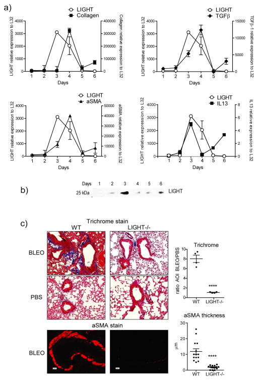
Background: Pulmonary fibrosis is characterized by excessive accumulation of collagen and α-smooth muscle actin in the lung. The key molecules that promote these phenotypes are of clinical interest.

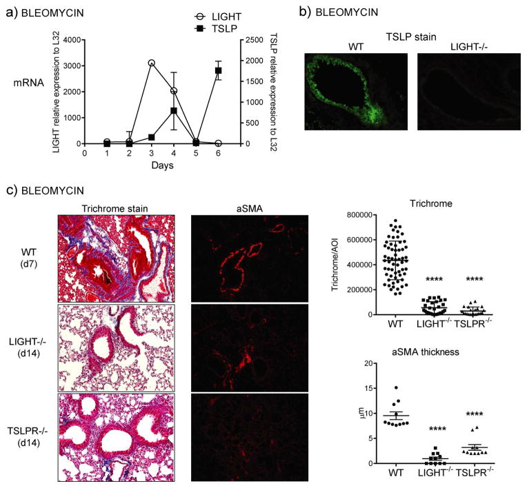
Objectives: Thymic stromal lymphopoietin (TSLP) has been found at high levels in patients with asthma and idiopathic pulmonary fibrosis, and TSLP has been proposed as a primary driver of lung fibrotic disease. We asked whether tumor necrosis factor superfamily protein 14 (TNFSF14) (aka LIGHT) controls TSLP production to initiate fibrosis.

Methods: Expression of TSLP and initiation of pulmonary fibrosis induced by bleomycin were assessed in mice deficient in LIGHT. The ability of recombinant LIGHT, given intratracheally to naive mice, to promote TSLP and fibrosis was also determined.

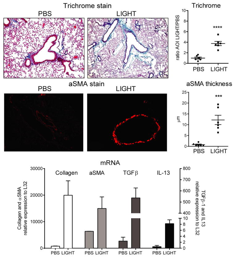
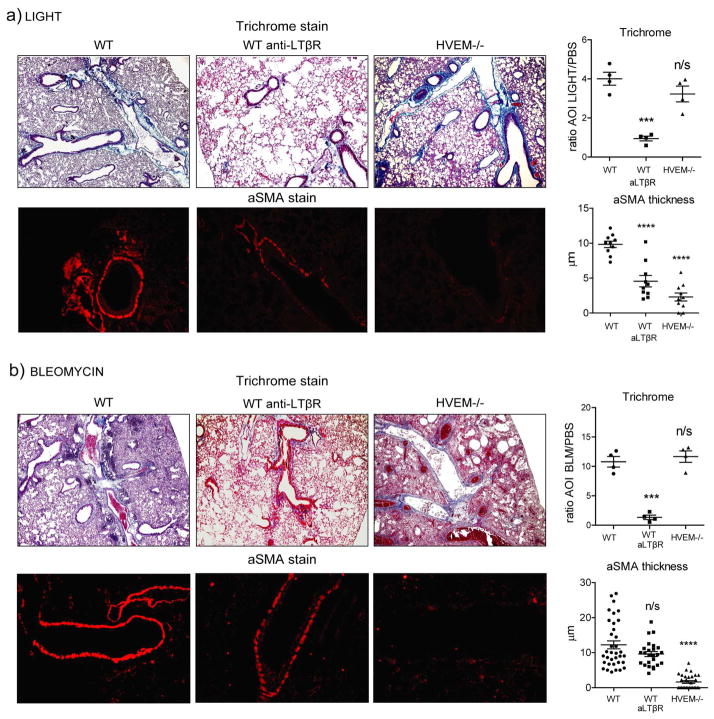
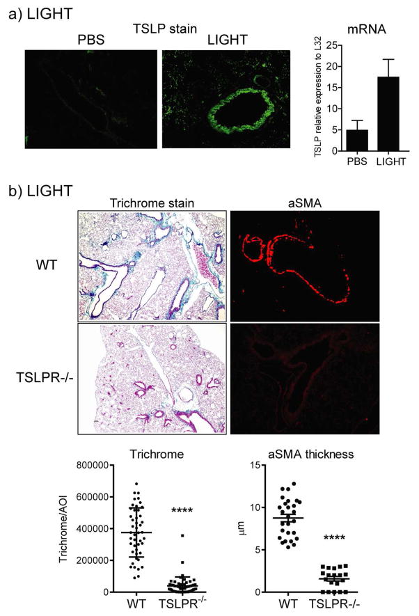
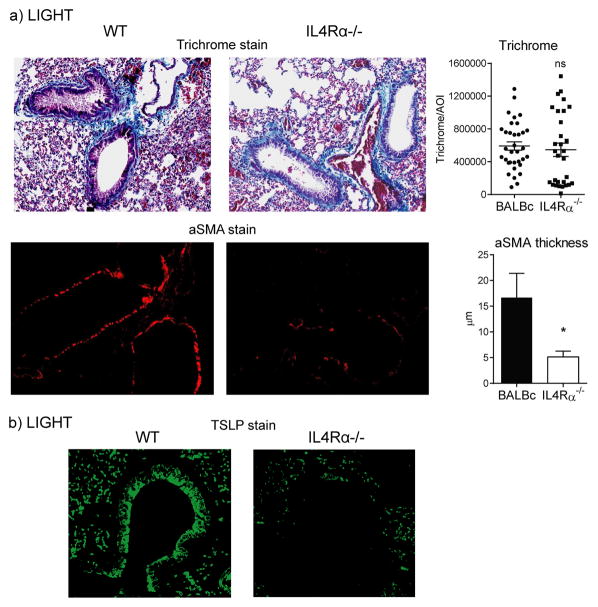
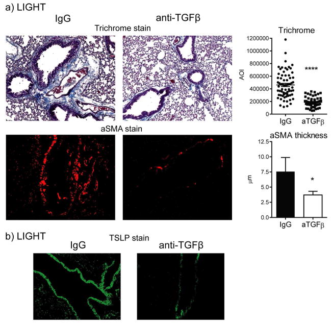
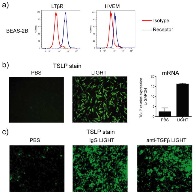
Results: Genetic deletion of LIGHT abolished lung TSLP expression driven by bleomycin, accompanied by near-complete absence of accumulation of lung collagen and α-smooth muscle actin. Furthermore, recombinant LIGHT administered in vivo induced lung expression of TSLP in the absence of other inflammatory stimuli, and strikingly reproduced the primary features of bleomycin-driven disease in a TSLP-dependent manner. Blockade of LIGHT binding to either of its receptors, herpes virus entry mediator and lymphotoxin beta receptor, inhibited clinical symptoms of pulmonary fibrosis, and correspondingly both receptors were found on human bronchial epithelial cells, a primary source of TSLP. Moreover, LIGHT induced TSLP directly in human bronchial epithelial cells and synergized with IL-13 and TGF-β in vivo to promote TSLP in the lungs and drive fibrosis.

Conclusions: These results show that LIGHT is a profibrogenic cytokine that may be a key driver of TSLP production during the initiation and development of lung fibrotic disease.

Keywords: HVEM; IPF; LIGHT; Pulmonary fibrosis; TNFSF14; TSLP; asthma; bronchial epithelial cell.


Figures

Figure 1
Figure 1. LIGHT is induced by bleomycin and LIGHT-deficient mice exhibit decreased lung fibrosis
(a–b) Wild type (WT) mice were sensitized with bleomycin intratracheally (i.t.). (a) Kinetics of expression of mRNA for LIGHT, alpha smooth muscle actin (aSMA), TGF-β, and IL-13, assessed by qPCR analysis of lung samples. mRNA expression calculated relative to L32. Values are mean ± SEM of 2 mice per time point. Selected time points were repeated in 3 additional experiments. (b) Soluble LIGHT was assessed in the bronchoalveolar lavage by Western Blot. Data are representative of 3 experiments. (c) WT and LIGHT−/− mice were sensitized with bleomycin or PBS intratracheally. Mice were sacrificed on day 7. Fibrotic phenotypes were assessed by analyzing collagen deposition (trichrome stain) and peribronchial aSMA accumulation in lung sections. Representative sections are shown (left). Quantitation was performed as in materials and methods (right). Trichrome data show mean ratio of area of interest (AOI) values over PBS of 4 individual mice per condition. aSMA data show values of 14 random bronchi scored from 4 mice per condition. Data are representative of 6 experiments.
Figure 2
Figure 2. Recombinant LIGHT induces lung fibrosis
WT mice were injected with 10 μg of soluble rmLIGHT administered intratracheally on days 1 and 2, and sacrificed on day 3. Fibrotic phenotypes were scored after analyzing trichrome stained and aSMA stained lung sections. Trichrome data show mean ratio of AOI values over PBS of 6 individual mice per condition. aSMA data show mean values of random bronchi scored from 6 mice per condition. qPCR analysis was performed on lung samples and mRNA expression of LIGHT, collagen, IL-13 and TGF-β calculated relative to L32. mRNA values are mean ± SEM of 3 mice per condition. Data are representative of 6 experiments.
Figure 3
Figure 3. HVEM and LTβR participate in promoting lung fibrotic activity induced by bleomycin and recombinant LIGHT
WT mice were compared to mice treated with 200 μg neutralizing anti-LTβR and to HVEM−/− mice. (a) Mice were treated with 10μg rmLIGHT intratracheally on days 1 and 2 before sacrificing on day 3. (b) Mice were treated with bleomycin intratracheally and sacrificed on day 7. Fibrotic phenotypes were assessed as before in the lungs. Trichrome data show mean ratio of AOI values over PBS of 4 individual mice per condition. aSMA data show values of 10 (a) to 35 (b) random bronchi scored from 4 mice per condition. Results are representative of 3 experiments performed.
Figure 4
Figure 4. TSLP expression in response to bleomycin is dependent on LIGHT
(a) Kinetics of LIGHT and TSLP mRNA expression by qPCR in lung samples of WT mice treated i.t. with bleomycin. (b) Lungs from WT and LIGHT−/− mice sensitized i.t. with bleomycin were assessed for TSLP expression on day 7 by IHC (green stain). (c) WT, LIGHT−/− and TSLPR−/− mice were compared for lung fibrotic phenotypes after sensitization i.t. with bleomycin. Trichrome and aSMA data show values of between 30–60 random AOI and 11 random bronchi, respectively, scored from 3 mice per group. Results are representative of 3 experiments.
Figure 5
Figure 5. Recombinant LIGHT induces TSLP expression in vivo to promote lung fibrosis
(a) TSLP expression was measured in the lungs by IHC or PCR 24h after the last LIGHT injection in WT mice treated with 10μg of rmLIGHT or PBS given i.t. on days 1 and 2. (b) WT and TSLPR−/− mice were treated with rmLIGHT as above and fibrotic activity in the lungs assessed on day 3. Trichrome and aSMA data show values of between 25–40 random AOI and 25 random bronchi, respectively, scored from 4 mice per group. Results are representative of 3 experiments.
Figure 6
Figure 6. LIGHT-induced lung fibrosis is partially dependent on IL-13 signaling
WT and IL-4Rα−/− mice were injected i.t. with rmLIGHT as before. Lung accumulation of collagen and aSMA (a), as well as TSLP expression by IHC (b), were assessed at day 3. Trichrome and aSMA data represent scores of between 30–40 random AOI and 30 random bronchi, respectively, from 3 individual mice per group.
Figure 7
Figure 7. TGF-β participates in lung fibrosis induced by LIGHT
WT mice were treated with anti-TGF-β or control IgG and injected i.t. with rmLIGHT as before. Lung accumulation of collagen and aSMA (a), as well as TSLP expression by IHC (b), were assessed at day 3. Trichrome and aSMA data represent scores of between 50–70 random AOI and 30 random bronchi, respectively, from 3 individual mice per group.
Figure 8
Figure 8. LIGHT directly induces TSLP expression in human bronchial epithelial cells in a TGF-β-independent manner
(a) Human bronchial epithelial cells (BEAS-2B) were assessed for LTβR and HVEM expression by flow cytometry. Isotype control in red. (b and c) BEAS-2B cells were stimulated in vitro with soluble rhLIGHT for 72h, in the absence (b) or presence of anti-TGF-β or control IgG (c). TSLP expression was evaluated by IHC and mRNA levels by qPCR. PCR values are mean ± SEM of triplicate samples per condition. Data are representative of 2–4 experiments.

References

    1. Thannickal VJ, Toews GB, White eS, Lynch P, Jr, Martinez FJ. Mechanisms of pulmonary fibrosis. Annu Rev Med. 2004;55:395–417. - PubMed
    1. Wilson MS, Wynn TA. Pulmonary fibrosis: pathogenesis, etiology and regulation. Mucosal Immunol. 2009 Jan 7;2(2):103–21. - PMC - PubMed
    1. Rosenbloom J, Castro SV, Jimenez SA. Narrative review: fibrotic diseases: cellular and molecular mechanisms and novel therapies. Ann Intern Med. 2010 Feb 2;152(3):159–66. [Research Support, Non-U.S. Gov’t Review] - PubMed
    1. Wynn TA, Ramalingam TR. Mechanisms of fibrosis: therapeutic translation for fibrotic disease. Nat Med. 2012 Jul;18(7):1028–40. [Research Support, N.I.H., Intramural Review] - PMC - PubMed
    1. Boin F, Wigley F. Connective tissue diseases: Immunosuppressive therapy in SSc: what is the target? Nat Rev Rheumatol. 2009 Jul;5(7):357–8. - PubMed

Publication types

MeSH terms