. 2015 Apr;166(4):1048-54.e1-5.
doi: 10.1016/j.jpeds.2014.12.069. Epub 2015 Feb 11.

Lymphatic and other vascular malformative/overgrowth disorders are caused by somatic mutations in PIK3CA

Affiliations

Lymphatic and other vascular malformative/overgrowth disorders are caused by somatic mutations in PIK3CA

Valerie L Luks et al. J Pediatr. 2015 Apr.

Abstract

Objectives: To test the hypothesis that somatic phosphatidylinositol-4,5-bisphospate 3-kinase, catalytic subunit alpha (PIK3CA) mutations would be found in patients with more common disorders including isolated lymphatic malformation (LM) and Klippel-Trenaunay syndrome (KTS).

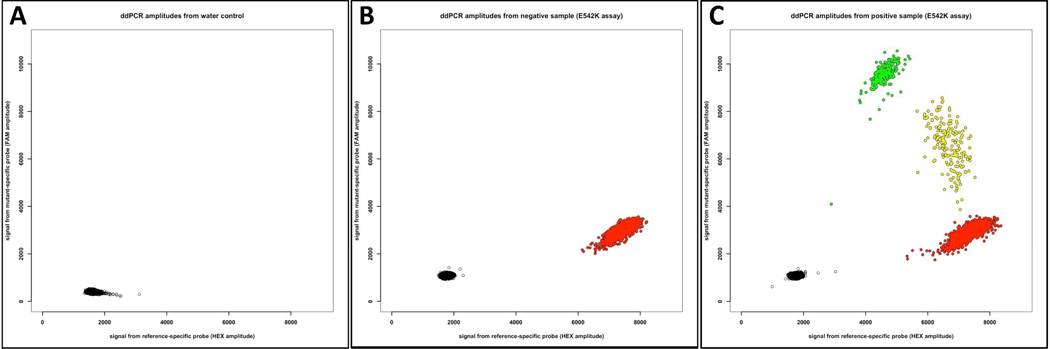
Study design: We used next generation sequencing, droplet digital polymerase chain reaction, and single molecule molecular inversion probes to search for somatic PIK3CA mutations in affected tissue from patients seen at Boston Children's Hospital who had an isolated LM (n = 17), KTS (n = 21), fibro-adipose vascular anomaly (n = 8), or congenital lipomatous overgrowth with vascular, epidermal, and skeletal anomalies syndrome (n = 33), the disorder for which we first identified somatic PIK3CA mutations. We also screened 5 of the more common PIK3CA mutations in a second cohort of patients with LM (n = 31) from Seattle Children's Hospital.

Results: Most individuals from Boston Children's Hospital who had isolated LM (16/17) or LM as part of a syndrome, such as KTS (19/21), fibro-adipose vascular anomaly (5/8), and congenital lipomatous overgrowth with vascular, epidermal, and skeletal anomalies syndrome (31/33) were somatic mosaic for PIK3CA mutations, with 5 specific PIK3CA mutations accounting for ∼ 80% of cases. Seventy-four percent of patients with LM from Seattle Children's Hospital also were somatic mosaic for 1 of 5 specific PIK3CA mutations. Many affected tissue specimens from both cohorts contained fewer than 10% mutant cells.

Conclusions: Somatic PIK3CA mutations are the most common cause of isolated LMs and disorders in which LM is a component feature. Five PIK3CA mutations account for most cases. The search for causal mutations requires sampling of affected tissues and techniques that are capable of detecting low-level somatic mosaicism because the abundance of mutant cells in a malformed tissue can be low.


Conflict of interest statement

The authors declare no conflicts of interest.

Figures

Figure 1
Figure 1
Photographs and MRIs of participants with isolated LM, CLOVES, KTS, and FAVA. (A) 8-month-old boy (LM1) with isolated LM. Note swelling in deltoid region without cutaneous vascular signs. Coronal and sagittal fat-saturated T2-weighted MRI demonstrates macrocystic LM (a multilocular cystic mass) involving the anterolateral aspects of the right shoulder without muscular infiltration (arrows); humeral head (asterisk). (B) 19-month-old female (CL12) with CLOVES syndrome. Note asymmetric distribution of truncal lipomatous masses and bilateral lower extremity involvement. Coronal fat-saturated T1-weighted MRI following contrast administration demonstrates moderate heterogeneous enhancement of the bilateral truncal masses (arrows). Axial T1-weighted MRI without contrast depicts truncal lipomatous overgrowth (arrows); segment VI of the liver (asterisk). (C) 3-year-old boy (KT4) with KTS. Note capillary-lymphatic malformation and overgrowth involving right lower extremity. Coronal and axial fat-saturated T2-weighted MRI shows the persistent marginal vein system (bent arrows) and marked enlargement of the subcutaneous tissues due to a combination of lymphatic fluid and fat (straight arrow). There are also intramuscular venous malformations. (D) 9 - year-old boy (F8) with FAVA of the left gastrocnemius muscle; note absence of overgrowth and cutaneous vascular anomalies. Sagittal fat-saturated T1-weighted MRI following contrast administration demonstrates the longitudinal distribution of the diffuse, fibro-adipose vascular anomaly (arrows). Axial fat-saturated T2-weighted MRI with (upper) and without (lower) contrast. Note right head of the gastrocnemius muscle is diffusely replaced by a contrast enhancing heterogeneous soft tissue lesion (arrows).
Figure 2
Figure 2
Scatterplots depicting ddPCR results specifically for the p.E542K mutation assay. The X-axis indicates fluorescence intensity in droplets for the probe targeting the wild-type amplimer and the Y-axis indicates fluorescence intensity in droplets for the probe targeting the p. E542K mutant amplimer. Individual droplets that contain no amplimer are pseudocolored black, wild-type amplimer red, p.E542K mutant amplimer green, and wild-type and p.E542K mutant amplimers yellow. (A) Scatterplot when water-alone was used as template. Neither wild-type nor mutant amplimer was present in any of the 15,633 droplets in this experiment. (B) Plot when affected tissue DNA from participant LM2, who has a different PIK3CA mutation (p.C420R), was used as template for the p.E542K assay. Of the 13,932 droplets analyzed, 11,670 contain no amplimer and 2,262 contain amplimer that is wild-type at the p.E542K locus. (C) Scatterplot when affected tissue DNA from participant LM1, who was previously found to have a p.E542K mutation by whole exome sequencing, was used as template. Of 13,146 droplets analyzed, 3,811 contain wild-type amplimer, 242 contain mutant amplimer, 193 contain wild-type and mutant amplimer, and 8,900 contain no amplimer.

Similar articles

Cited by

References

    1. Samuels Y, Wang Z, Bardelli A, Silliman N, Ptak J, Szabo S, et al. High frequency of mutations of the PIK3CA gene in human cancers. Science. 2004;304:554. - PubMed
    1. Zhao L, Vogt PK. Class I PI3K in oncogenic cellular transformation. Oncogene. 2008;27:5486–5496. - PMC - PubMed
    1. Kinross KM, Montgomery KG, Kleinschmidt M, Waring P, Ivetac I, Tikoo A, et al. An activating Pik3ca mutation coupled with Pten loss is sufficient to initiate ovarian tumorigenesis in mice. J Clin Invest. 2012;122:553–557. - PMC - PubMed
    1. Kurek KC, Luks VL, Ayturk UM, Alomari AI, Fishman SJ, Spencer SA, et al. Somatic mosaic activating mutations in PIK3CA cause CLOVES syndrome. Am J Hum Genet. 2012;90:1108–1115. - PMC - PubMed
    1. Lee JH, Huynh M, Silhavy JL, Kim S, Dixon-Salazar T, Heiberg A, et al. De novo somatic mutations in components of the PI3K-AKT3-mTOR pathway cause hemimegalencephaly. Nature genetics. 2012;44:941–945. - PMC - PubMed

Publication types

MeSH terms