

Posttranscriptional deregulation of signaling pathways in meningioma subtypes by differential expression of miRNAs
- PMID: 25681310
- PMCID: PMC4588755
- DOI: 10.1093/neuonc/nov014
Posttranscriptional deregulation of signaling pathways in meningioma subtypes by differential expression of miRNAs
Abstract
Background: Micro (mi)RNAs are key regulators of gene expression and offer themselves as biomarkers for cancer development and progression. Meningioma is one of the most frequent primary intracranial tumors. As of yet, there are limited data on the role of miRNAs in meningioma of different histological subtypes and the affected signaling pathways.
Methods: In this study, we compared expression of 1205 miRNAs in different meningioma grades and histological subtypes using microarrays and independently validated deregulation of selected miRNAs with quantitative real-time PCR. Clinical utility of a subset of miRNAs as biomarkers for World Health Organization (WHO) grade II meningioma based on quantitative real-time data was tested. Potential targets of deregulated miRNAs were discovered with an in silico analysis.
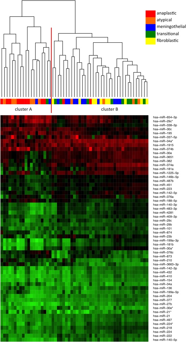
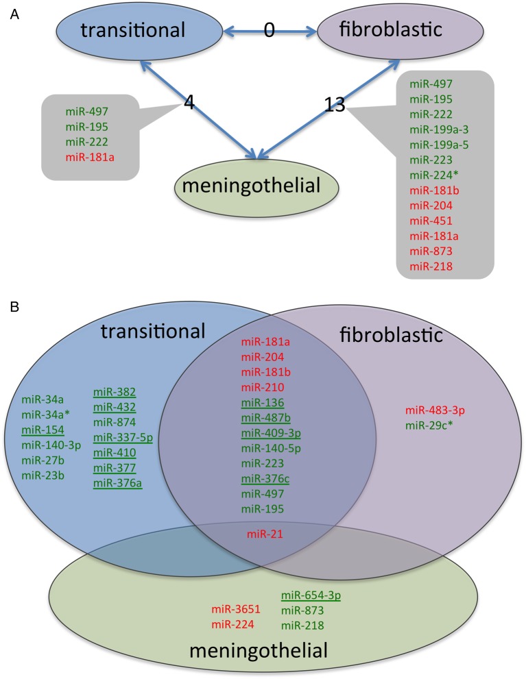
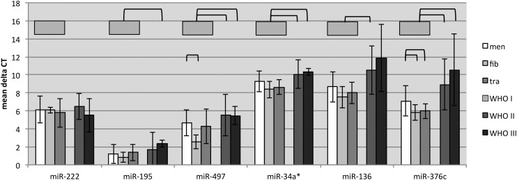
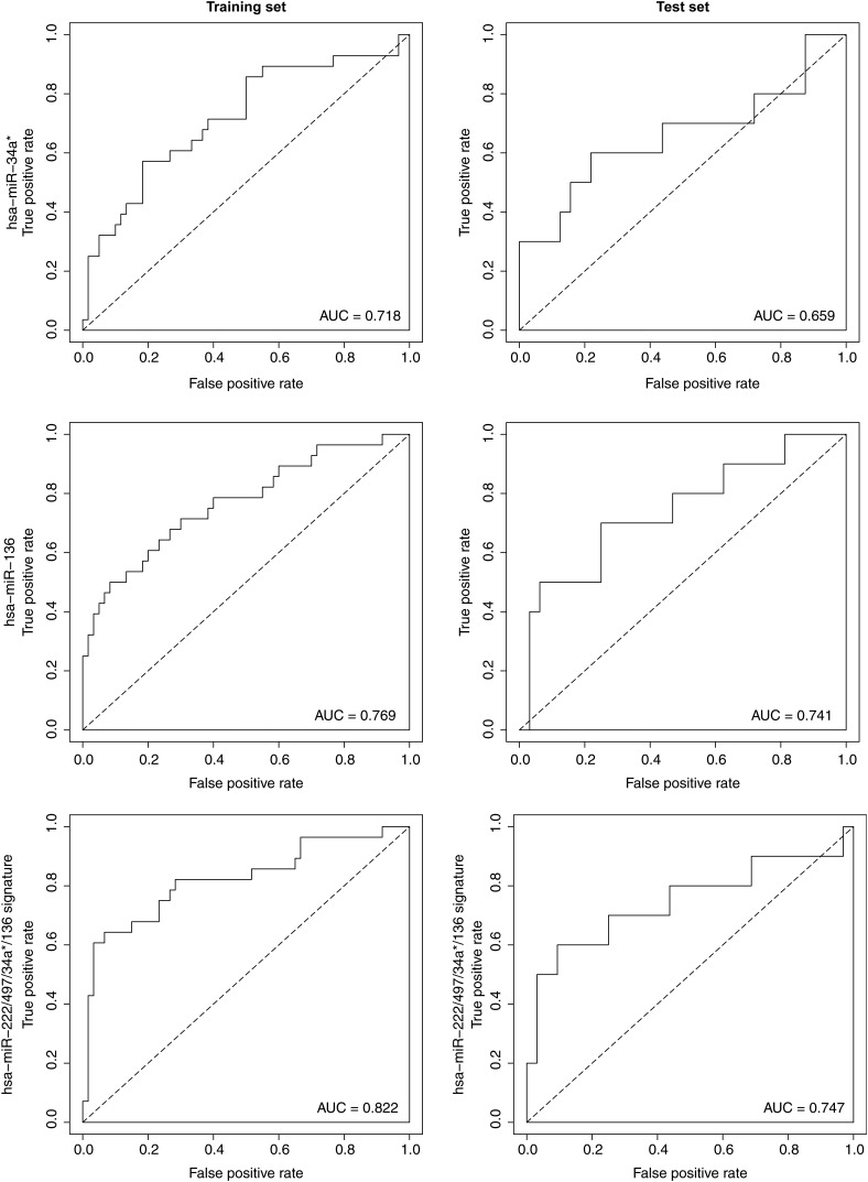
Results: We identified 13 miRNAs deregulated between different subtypes of benign meningiomas, and 52 miRNAs deregulated in anaplastic meningioma compared with benign meningiomas. Known and putative target genes of deregulated miRNAs include genes involved in epithelial-to-mesenchymal transition for benign meningiomas, and Wnt, transforming growth factor-β, and vascular endothelial growth factor signaling for higher-grade meningiomas. Furthermore, a 4-miRNA signature (miR-222, -34a*, -136, and -497) shows promise as a biomarker differentiating WHO grade II from grade I meningiomas with an area under the curve of 0.75.
Conclusions: Our data provide novel insights into the contribution of miRNAs to the phenotypic spectrum in benign meningiomas. By deregulating translation of genes belonging to signaling pathways known to be important for meningioma genesis and progression, miRNAs provide a second in line amplification of growth promoting cellular signals. MiRNAs as biomarkers for diagnosis of aggressive meningiomas might prove useful and should be explored further in a prospective manner.
Keywords: histological subtypes; meningioma; miRNA; qRT-PCR.
© The Author(s) 2015. Published by Oxford University Press on behalf of the Society for Neuro-Oncology. All rights reserved. For permissions, please e-mail: journals.permissions@oup.com.
Figures
References
-
- Bartel DP. MicroRNAs: genomics, biogenesis, mechanism, and function. Cell. 2004;116(2):281–297. - PubMed
-
- Perry ALD, Scheithauer BW, Budka H, et al. Meningiomas. In: Louis DNOH, Wiestler OD, Cavenee WK, eds. WHO Classification of Tumors of the Central Nervous System. Lyon: IARC press; 2007: 164–172.
-
- Stessin AM, Schwartz A, Judanin G, et al. Does adjuvant external-beam radiotherapy improve outcomes for nonbenign meningiomas? A Surveillance, Epidemiology, and End Results (SEER)–based analysis. J Neurosurg. 2012;117(4):669–675. - PubMed
-
- Perry A, Gutmann DH, Reifenberger G. Molecular pathogenesis of meningiomas. J Neurooncol. 2004;70(2):183–202. - PubMed
Publication types
MeSH terms
Substances
Associated data
- Actions
LinkOut - more resources
Full Text Sources
Other Literature Sources
Molecular Biology Databases
Research Materials
