Review
. 2015 May;64(5):830-41.
doi: 10.1136/gutjnl-2014-306842. Epub 2015 Feb 13.

Pathobiology of liver fibrosis: a translational success story

Affiliations
Review

Pathobiology of liver fibrosis: a translational success story

Youngmin A Lee et al. Gut. 2015 May.

Erratum in

  • Correction.
    [No authors listed] [No authors listed] Gut. 2015 Aug;64(8):1337. doi: 10.1136/gutjnl-2014-306842corr1. Gut. 2015. PMID: 26160826 No abstract available.

Abstract

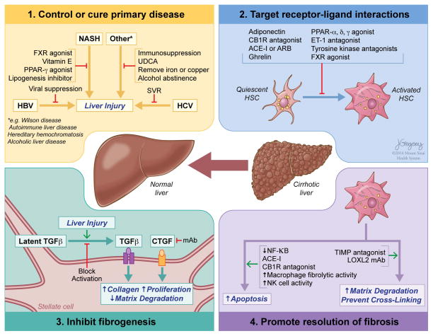
Reversibility of hepatic fibrosis and cirrhosis following antiviral therapy for hepatitis B or C has advanced the prospect of developing antifibrotic therapies for patients with chronic liver diseases, especially non-alcoholic steatohepatitis. Mechanisms of fibrosis have focused on hepatic stellate cells, which become fibrogenic myofibroblasts during injury through 'activation', and are at the nexus of efforts to define novel drug targets. Recent studies have clarified pathways of stellate cell gene regulation and epigenetics, emerging pathways of fibrosis regression through the recruitment and amplification of fibrolytic macrophages, nuanced responses of discrete inflammatory cell subsets and the identification of the 'ductular reaction' as a marker of severe injury and repair. Based on our expanded knowledge of fibrosis pathogenesis, attention is now directed towards strategies for antifibrotic therapies and regulatory challenges for conducting clinical trials with these agents. New therapies are attempting to: 1) Control or cure the primary disease or reduce tissue injury; 2) Target receptor-ligand interactions and intracellular signaling; 3) Inhibit fibrogenesis; and 4) Promote resolution of fibrosis. Progress is urgently needed in validating non-invasive markers of fibrosis progression and regression that can supplant biopsy and shorten the duration of clinical trials. Both scientific and clinical challenges remain, however the past three decades of steady progress in understanding liver fibrosis have contributed to an emerging translational success story, with realistic hopes for antifibrotic therapies to treat patients with chronic liver disease in the near future.

Keywords: CIRRHOSIS; EXTRACELLULAR MATRIX; FATTY LIVER; HEPATIC FIBROSIS; HEPATIC STELLATE CELL.


Conflict of interest statement

Competing interests: None.

Figures

Figure 1
Figure 1
Functions, features and phenotypes of hepatic stellate cells (HSCs) in normal and diseased liver. HSCs may exist as several different phenotypes with distinct molecular and cellular functions and features, each of which contributes significantly to liver homeostasis and disease. Quiescent stellate cells are critical to the normal metabolic functioning of the liver. Liver injury provokes the transdifferentiation of quiescent stellate cells to their activated phenotype, leading to metabolic reprogramming, increased autophagy to fuel the metabolic demands, amplification of parenchymal injury and the development of ‘classic’ phenotypic features of activated HSCs/myofibroblasts. Through these changes, activated stellate cells drive the fibrotic response to injury and the development of cirrhosis. As liver injury subsides, activated stellate cells can be eliminated by one of three pathways: apoptosis, senescence or reversion to an inactivated phenotype. Senescent stellate cells are more likely to be cleared by NK cell-mediated cell death while inactivated stellate cells remain ‘primed’ to respond to further liver injury. This reduction in the number of activated stellate cells contributes to the regression of fibrosis or cirrhosis and repair of the liver in most, but not all patients. The relative contribution of these three pathways of stellate cell clearance to fibrosis regression is not yet clear. ECM, extracellular matrix; HSCs, hepatic stellate cells. NK, natural killer.
Figure 2
Figure 2
Extrahepatic factors that affect liver fibrosis. In addition to intrahepatic injury signals, extrahepatic factors are increasingly recognised to drive liver fibrosis. Intestinal dysbiosis and bacterial overgrowth contribute to a ‘fibrogenic microbiome’, especially in cholestatic liver diseases and non-alcoholic steatohepatitis (NASH). Enterohepatic circulation of bile acids mediates bacteriostasis and promotes protection from hepatic fibrosis through increased farnesoid-X-receptor (FXR)/TGR5 signalling. Proinflammatory signalling (TNFα, IL-6) and adipokines secreted from adipose tissue mediate profibrogenic (eg, leptin, resistin) or protective (eg, adiponectin) effects on liver. Insulin resistance and metabolic syndrome are risk factors for progression in chronic liver diseases (eg, NASH, HCV). Hyperinsulinemia promotes steatosis, the generation of reactive oxygen species and lipid peroxides. Vascular abnormalities may also contribute to the development of hepatic fibrosis. The interactions that promote liver fibrosis are depicted with green lines and protective interactions with red lines.
Figure 3
Figure 3
Epigenetic mechanisms and post transcriptional gene regulation of hepatic stellate cells. ‘Epigenetics’ is defined as heritable traits that are not linked to changes in DNA sequence, involving mechanisms by which chromatin-associated proteins and post-translational modifications of histones regulate transcription. MicroRNAs mediate post-transcriptional regulation by promoting mRNA degradation and translational repression. Abnormal patterns of DNA methylation identified in liver fibrosis and activated stellate cells include, for example, hypermethylation of Phosphatase and Tension Homologue (PTEN) with consequent gene repression. PTEN negatively regulates the activation of ERK and AKT signalling pathways controlling cell cycling, proliferation, focal adhesion and cell migration. Repression of PTEN in activated stellate cells thereby promotes fibrogenesis. Similarly, hypermethylation of, and gene repression by MeCP2 of RASAL1, IkB, PPAR-γ and PTCH1 lead to inhibition of ERK signalling pathways, or loss of inhibition of GLI1 and SMAD3, respectively, thus promoting hepatic stellate cell (HSC) survival (IkB), and HSC activation and fibrogenesis. Histone modifications with profibrotic effects have been identified in activated HSCs and include MRTF-A and TGFβ-dependent chromatin remodeling leading to the altered binding of vitamin D receptor and SMAD3 mediated transcription of fibrogenic genes. Examples of microRNAs promoting antifibrotic and profibrotic effects are shown.
Figure 4
Figure 4
Inflammatory and immune cell interactions that promote or inhibit the activation of hepatic stellate cells. Hepatic cells promoting (green lines) or inhibiting (red lines) the activation of quiescent stellate cells to activated, fibrogenic hepatic stellate cells are shown. Examples of common mediators of these responses are included. Hh, hedgehog ligands; NK, natural killer.
Figure 5
Figure 5
Mechanisms by which antifibrotic therapies may lead to fibrosis regression. (1) Disease-specific therapies that control or cure the underlying disease are still the most effective antifibrotic approach. (2) Targeting receptor–ligand interactions with either established or experimental drugs to reduce hepatic stellate cell activation will attenuate fibrosis development, with multiple potential strategies under development. (3) Inhibition of the most potent of the profibrogenic pathways, for example, preventing activation of latent TGFβ, or blocking the activity of CTGF, are among the more promising antifibrotic strategies. (4) The resolution of fibrosis can be promoted by enhancing the apoptosis of activated hepatic stellate cells either with drugs or through the activity of either NK cells and by increasing the degradation of extracellular matrix, by fibrolytic macrophages or preventing its cross-linking with antagonists to LOXL2. FXR, farnesoid-X-receptor; PPAR, peroxisome proliferator-activated receptor; UDCA, ursodeoxycholic acid; SVR, sustained virological response; CB1, cannabinoid receptor type 1; ARB, angiotensin II receptor blocker; ET-1, endothelin 1; TGFβ, transforming growth factor β; CTGF, connective tissue growth factor; mAb, monoclonal antibody; NF-κB, nuclear factor kappa-light-chain-enhancer of activated B cells; NK, natural killer; TIMP, tissue inhibitor of metalloproteinase; LOXL2, Lysyl oxidase 2.

Comment in

References

    1. Perez-Tamayo R. Cirrhosis of the liver: a reversible disease? Pathol Annu. 1979;14 (Pt 2):183–213. - PubMed
    1. Okazaki I, Maruyama K. Collagenase activity in experimental hepatic fibrosis. Nature. 1974;252:49–50. - PubMed
    1. Pellicoro A, Ramachandran P, Iredale JP, et al. Liver fibrosis and repair: immune regulation of wound healing in a solid organ. Nat Rev Immunol. 2014;14:181–94. - PubMed
    1. Marcellin P, Gane E, Buti M, et al. Regression of cirrhosis during treatment with tenofovir disoproxil fumarate for chronic hepatitis B: a 5-year open-label follow-up study. Lancet. 2013;381:468–75. - PubMed
    1. D’Ambrosio R, Aghemo A, Rumi MG, et al. A morphometric and immunohistochemical study to assess the benefit of a sustained virological response in hepatitis C virus patients with cirrhosis. Hepatology. 2012;56:532–43. - PubMed

Publication types

MeSH terms