. 2015 Apr;138(Pt 4):946-62.
doi: 10.1093/brain/awv018. Epub 2015 Feb 12.

Reduced cortical innervation of the subthalamic nucleus in MPTP-treated parkinsonian monkeys

Affiliations

Reduced cortical innervation of the subthalamic nucleus in MPTP-treated parkinsonian monkeys

Abraham Mathai et al. Brain. 2015 Apr.

Abstract

The striatum and the subthalamic nucleus are the main entry points for cortical information to the basal ganglia. Parkinson's disease affects not only the function, but also the morphological integrity of some of these inputs and their synaptic targets in the basal ganglia. Significant morphological changes in the cortico-striatal system have already been recognized in patients with Parkinson's disease and in animal models of the disease. To find out whether the primate cortico-subthalamic system is also subject to functionally relevant morphological alterations in parkinsonism, we used a combination of light and electron microscopy anatomical approaches and in vivo electrophysiological methods in monkeys rendered parkinsonian following chronic exposure to low doses of 1-methyl-4-phenyl-1,2,3,6-tetrahydropyridine (MPTP). At the light microscopic level, the density of vesicular glutamate transporter 1-positive (i.e. cortico-subthalamic) profiles in the dorsolateral part of the subthalamic nucleus (i.e. its sensorimotor territory) was 26.1% lower in MPTP-treated parkinsonian monkeys than in controls. These results were confirmed by electron microscopy studies showing that the number of vesicular glutamate transporter 1-positive terminals and of axon terminals forming asymmetric synapses in the dorsolateral subthalamic nucleus was reduced by 55.1% and 27.9%, respectively, compared with controls. These anatomical findings were in line with in vivo electrophysiology data showing a 60% reduction in the proportion of pallidal neurons that responded to electrical stimulation of the cortico-subthalamic system in parkinsonian monkeys. These findings provide strong evidence for a partial loss of the hyperdirect cortico-subthalamic projection in MPTP-treated parkinsonian monkeys.

Keywords: Parkinson’s disease; cortico-subthalamic; hyperdirect; synaptic plasticity; vesicular glutamate transporter.


Figures

Figure 1
Figure 1
Extent of dopaminergic denervation in MPTP-treated monkeys. (A–C) Light microscopic images of coronal brain sections showing immunostaining for tyrosine hydroxylase (TH) in the post-commissural putamen of a control animal (A) and the two MPTP-treated monkeys (B and C) that were used in the electrophysiological studies (Monkeys I and O). The pattern of tyrosine hydroxylase immunoreactivity in the striatum of the MPTP-treated animals used in the anatomical studies was comparable to that shown in Monkey O. Cd = caudate nucleus; IC = internal capsule; Put = putamen. Scale bars: A–C = 1 mm. (D) Normalized densitometry measurements of tyrosine hydroxylase immunoreactivity in the post-commissural putamen of normal (n = 2) and MPTP-treated (n = 8) monkeys. ***Significant difference from normal; P < 0.001; Student’s t-test).
Figure 2
Figure 2
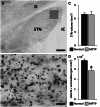
vGluT1 immunostaining in the monkey STN. (A) Light microscopic view of a coronal section through the monkey STN showing immunostaining for vGluT1. The grey square indicates the size and approximate position of the region of interest in the dorsolateral territory of the STN that was used to quantify the density of vGluT1-positive varicose processes. Scale bar = 0.5 mm. ZI = zona incerta; IC = internal capsule. (B) Light micrograph showing vGluT1-immunopositive pleomorphic varicose processes in the dorsolateral STN of a normal monkey. Scale bar = 10 µm. (C) Comparison of the average STN volume ( ± SEM; n = 3) between normal and parkinsonian monkeys. (D) Average density ( ± SEM; n = 3) of vGluT1-immunoreactive varicosities in the dorsolateral STN of normal and parkinsonian monkeys. *P = 0.012; Student’s t-test of difference between the normal and parkinsonian animals.
Figure 3
Figure 3
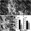
Changes in vGluT1-immunoreactive terminals density in the STN of parkinsonian monkeys. (A) Electron micrographs showing vGluT1-positive (v1-immunoperoxidase) and vGluT2-labelled (v2-immunogold) terminals in the dorsolateral STN. Scale bar = 0.5 µm. (B and C) Electron micrographs depicting an asymmetric synapse (putatively glutamatergic) in consecutive ultrathin serial sections of the dorsolateral monkey STN. Asymmetric synapse indicated by arrowheads. Scale bars: B and C = 0.2 µm. (D) Average density ( ± SEM; n = 3) of vGluT1-immunopositive terminals in the dorsolateral STN of normal and parkinsonian monkeys. *P = 0.02; Student’s t-test of difference between normal and parkinsonian animals. Total surface area analysed: 3960 µm2/animal. (E) Average density ( ± SEM; n = 3) of asymmetric synapses in the dorsolateral STN of normal and parkinsonian monkeys. *P = 0.029; Student’s t-test of difference between normal and parkinsonian animals. Total surface area analysed: 1980 µm2/animal.
Figure 4
Figure 4
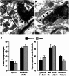
Post-synaptic targets of vGluT1-immunopositive terminals in the dorsolateral STN. (A and B) Electron micrographs showing vGluT1-containing terminals (v1) forming asymmetric synapses (indicated by arrowheads) with a dendritic shaft (dend) (A) and a dendritic spine (sp) (B). The black line with double-ended arrows on the dendritic shaft (A) illustrates the measurement of its cross-sectional diameter, defined as the shortest diameter passing through its centre. Scale bars: A and B = 0.2 µm. (C) Post-synaptic targets of vGluT1-immunopositive terminals in the dorsolateral STN. No significant difference was found in the proportion of vGluT1-immunoreactive terminals forming asymmetric synapses with dendritic shafts and spines in normal versus parkinsonian monkeys (left). The right panel shows the size distribution of all postsynaptic dendritic shafts contacted by vGluT1-immunoreactive terminals, which is not significantly different between normal and parkinsonian animals. Lg = large; Md = medium; Sm = small. Columns represent means ± SEM across three normal and three parkinsonian monkeys.
Figure 5
Figure 5
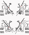
Approximate locations of the placements of the stimulation electrodes projected onto coronal representations of the monkey brain. Experiments in the normal condition were conducted in the left hemisphere, whereas those in the parkinsonian states were performed on the right side. Thick black lines indicate the paths of the stimulation electrode with the red tips marking the positions of the electrical contact points of the electrode. Antero-posterior coordinates were determined by comparing the brain sections with a standard monkey atlas (Paxinos et al., 1999). Put = putamen; Cd = caudate; Thal = thalamus; Ctx = cerebral cortex; SN = substantia nigra.
Figure 6
Figure 6
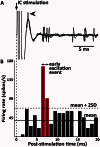
Response of a pallidal neuron to internal capsule (IC) stimulation. (A) Six overlaid traces of neuronal activity show that the neuron often fires around 6–8 ms post-stimulation. The arrowhead points to a neuronal spike occurring along with the stimulation artefact. The neuronal spike was successfully recognized after processing the data with a stimulus artefact removal algorithm. (B) Peri-stimulus histogram depicting the firing rate of the neuron during the first 20 ms post-stimulation shows an early excitation response (red) to internal capsule stimulation. Peri-stimulus histogram generated from 100 randomly spaced consecutive stimuli (bin size = 1 ms), as described in the ‘Materials and methods’ section. The neuronal traces (A) and peri-stimulus histograms (B) are aligned to identical time axes.
Figure 7
Figure 7
Electrophysiological responses of pallidal neurons to internal capsule stimulation. (A) Proportion of pallidal neurons responding with a characteristic early excitation to internal capsule stimulation in normal and parkinsonian monkeys. *P = 0.046 (Student’s t-test of difference between normal and parkinsonian state). Means ± SEM across two animals. (B) Intensity of early excitatory events in pallidal neurons showing an early excitatory response in normal and parkinsonian states (see ‘Materials and methods’ section for details). (C) Latency of early excitation responses in pallidal neurons in the normal and parkinsonian conditions. (D) Percent failure rate of spike occurrence during the early excitation response epoch after individual stimuli. Columns B–D represent means ± SEM across neurons with an early excitation response.

References

    1. Baudrexel S, Witte T, Seifried C, von Wegner F, Beissner F, Klein JC, et al. Resting state fMRI reveals increased subthalamic nucleus-motor cortex connectivity in Parkinson's disease. Neuroimage. 2011;55:1728–38. - PubMed
    1. Baufreton J, Atherton JF, Surmeier DJ, Bevan MD. Enhancement of excitatory synaptic integration by GABAergic inhibition in the subthalamic nucleus. J Neurosci. 2005;25:8505–17. - PMC - PubMed
    1. Baufreton J, Bevan MD. D2-like dopamine receptor-mediated modulation of activity-dependent plasticity at GABAergic synapses in the subthalamic nucleus. J Physiol. 2008;586:2121–42. - PMC - PubMed
    1. Bergman H, Wichmann T, Karmon B, DeLong MR. The primate subthalamic nucleus. II. Neuronal activity in the MPTP model of parkinsonism. J Neurophysiol. 1994;72:507–20. - PubMed
    1. Bevan MD, Francis CM, Bolam JP. The glutamate-enriched cortical and thalamic input to neurons in the subthalamic nucleus of the rat: convergence with GABA-positive terminals. J Comp Neurol. 1995;361:491–511. - PubMed

Publication types

MeSH terms

Substances