

Environmentally persistent free radicals compromise left ventricular function during ischemia/reperfusion injury
- PMID: 25681431
- PMCID: PMC4551132
- DOI: 10.1152/ajpheart.00891.2014
Environmentally persistent free radicals compromise left ventricular function during ischemia/reperfusion injury
Abstract
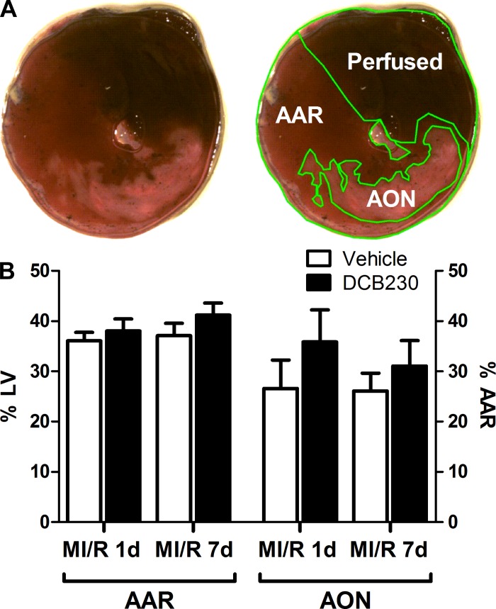
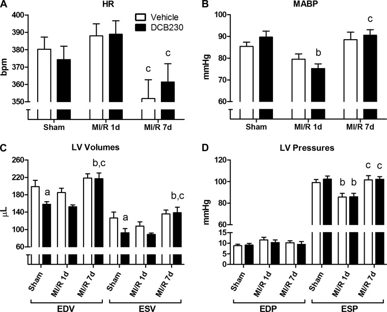
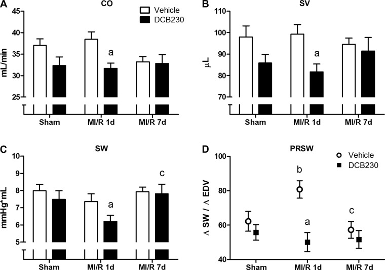
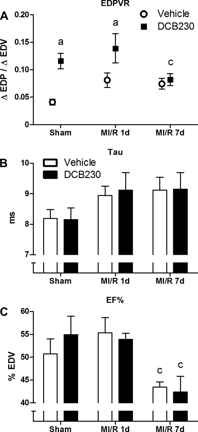
Increases in airborne particulate matter (PM) are linked to increased mortality from myocardial ischemia. PM contains environmentally persistent free radicals (EPFRs) that form as halogenated hydrocarbons chemisorb to transition metal oxide-coated particles, and are capable of sustained redox cycling. We hypothesized that exposure to the EPFR DCB230 would increase cardiac vulnerability to subsequent myocardial ischemia-reperfusion (MI/R) injury. Rats were exposed to DCB230 or vehicle via nose-only inhalation (230 μg max/day) over 30 min/day for 7 days. MI/R or sham MI/R (sham) was initiated 24 h after the final exposure. Following 1 or 7 days of reperfusion, left ventricular (LV) function was assessed and infarct size measured. In vehicle-exposed rats, MI/R injury did not significantly reduce cardiac output (CO), stroke volume (SV), stroke work (SW), end-diastolic volume (EDV), or end-systolic volume (ESV) after 1 day of reperfusion, despite significant reductions in end-systolic pressure (ESP). Preload-recruitable SW (PRSW; contractility) was elevated, presumably to maintain LV function. MI/R 1-day rats exposed to DCB230 also had similarly reduced ESP. Compared with vehicle controls, CO, SV, and SW were significantly reduced in DCB230-exposed MI/R 1-day rats; moreover, PRSW did not increase. DCB230's effects on LV function dissipated within 8 days of exposure. These data show that inhalation of EPFRs can exacerbate the deficits in LV function produced by subsequent MI/R injury. Infarct size was not different between the MI/R groups. We conclude that inhalation of EPFRs can compromise cardiac function during MI/R injury and may help to explain the link between PM and MI/R-related mortality.
Keywords: cardiac toxicity; particulate matter; pressure volume; rat.
Copyright © 2015 the American Physiological Society.
Figures
Similar articles
-
Environmentally persistent free radicals decrease cardiac function and increase pulmonary artery pressure.Am J Physiol Heart Circ Physiol. 2012 Nov 1;303(9):H1135-42. doi: 10.1152/ajpheart.00545.2012. Epub 2012 Aug 31. Am J Physiol Heart Circ Physiol. 2012. PMID: 22942180 Free PMC article.
-
Environmentally persistent free radicals decrease cardiac function before and after ischemia/reperfusion injury in vivo.J Recept Signal Transduct Res. 2011 Apr;31(2):157-67. doi: 10.3109/10799893.2011.555767. J Recept Signal Transduct Res. 2011. PMID: 21385100 Free PMC article.
-
Infusion of Melatonin Into the Paraventricular Nucleus Ameliorates Myocardial Ischemia-Reperfusion Injury by Regulating Oxidative Stress and Inflammatory Cytokines.J Cardiovasc Pharmacol. 2019 Oct;74(4):336-347. doi: 10.1097/FJC.0000000000000711. J Cardiovasc Pharmacol. 2019. PMID: 31356536 Free PMC article.
-
Clinical aspects of left ventricular diastolic function assessed by Doppler echocardiography following acute myocardial infarction.Dan Med Bull. 2001 Nov;48(4):199-210. Dan Med Bull. 2001. PMID: 11767125 Review.
-
Pharmacological prevention of reperfusion injury in acute myocardial infarction. A potential role for adenosine as a therapeutic agent.Am J Cardiovasc Drugs. 2004;4(3):159-67. doi: 10.2165/00129784-200404030-00003. Am J Cardiovasc Drugs. 2004. PMID: 15134468 Review.
Cited by
-
Environmentally Persistent Free Radicals Cause Apoptosis in HL-1 Cardiomyocytes.Cardiovasc Toxicol. 2017 Apr;17(2):140-149. doi: 10.1007/s12012-016-9367-x. Cardiovasc Toxicol. 2017. PMID: 27052339 Free PMC article.
-
Ginsenoside Rg3 attenuates myocardial ischemia/reperfusion-induced ferroptosis via the keap1/Nrf2/GPX4 signaling pathway.BMC Complement Med Ther. 2024 Jun 26;24(1):247. doi: 10.1186/s12906-024-04492-4. BMC Complement Med Ther. 2024. PMID: 38926825 Free PMC article.
-
miR-21 enhances the protective effect of loperamide on rat cardiomyocytes against hypoxia/reoxygenation, reactive oxygen species production and apoptosis via regulating Akap8 and Bard1 expression.Exp Ther Med. 2019 Feb;17(2):1312-1320. doi: 10.3892/etm.2018.7047. Epub 2018 Dec 5. Exp Ther Med. 2019. PMID: 30680008 Free PMC article.
-
A critical review of environmentally persistent free radical (EPFR) solvent extraction methodology and retrieval efficiency.Chemosphere. 2021 Dec;284:131353. doi: 10.1016/j.chemosphere.2021.131353. Epub 2021 Jun 29. Chemosphere. 2021. PMID: 34225117 Free PMC article. Review.
-
Soil contamination by environmentally persistent free radicals and dioxins following train derailment in East Palestine, OH.Environ Sci Process Impacts. 2025 Mar 19;27(3):729-740. doi: 10.1039/d4em00609g. Environ Sci Process Impacts. 2025. PMID: 39981852 Free PMC article.
References
-
- Bagate K, Meiring JJ, Gerlofs-Nijland ME, Cassee FR, Wiegand H, Osornio-Vargas A, Borm a PJ. Ambient particulate matter affects cardiac recovery in a Langendorff ischemia model. Inhal Toxicol 18: 633–643, 2006. - PubMed
-
- Brook RD, Franklin B, Cascio W, Hong Y, Howard G, Lipsett M, Luepker R, Mittleman M, Samet J, Smith SC, Tager I. Air pollution and cardiovascular disease: a statement for healthcare professionals from the Expert Panel on Population and Prevention Science of the American Heart Association. Circulation 109: 2655–2671, 2004. - PubMed
-
- Brook RD, Rajagopalan S, Pope CA, Brook JR, Bhatnagar A, Diez-Roux AV, Holguin F, Hong Y, Luepker RV, Mittleman MA, Peters A, Siscovick D, Smith SC, Whitsel L, Kaufman JD. Particulate matter air pollution and cardiovascular disease: An update to the scientific statement from the American Heart Association. Circulation 121: 2331–2378, 2010. - PubMed
Publication types
MeSH terms
Substances
Grants and funding
LinkOut - more resources
Full Text Sources
Other Literature Sources
Medical