. 2015 Apr 3;290(14):9075-86.
doi: 10.1074/jbc.M114.619874. Epub 2015 Feb 13.

Subcellular localization and Ser-137 phosphorylation regulate tumor-suppressive activity of profilin-1

Affiliations

Subcellular localization and Ser-137 phosphorylation regulate tumor-suppressive activity of profilin-1

Marc I Diamond et al. J Biol Chem. .

Abstract

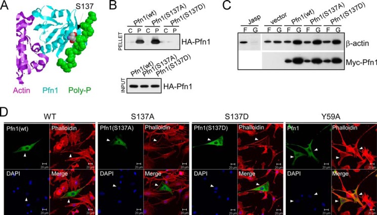
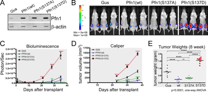
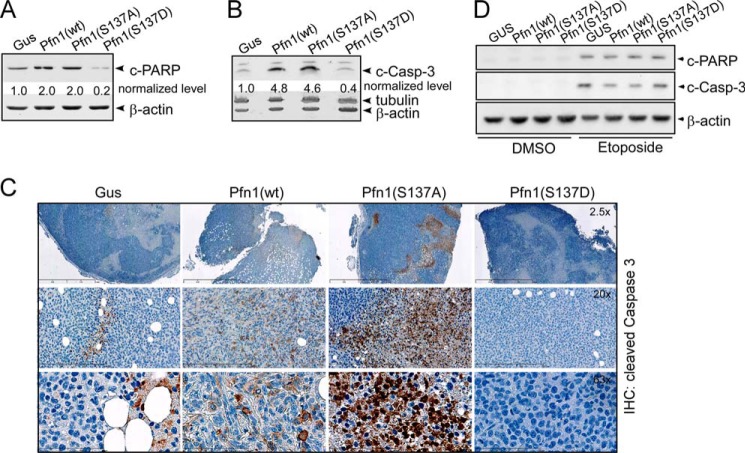
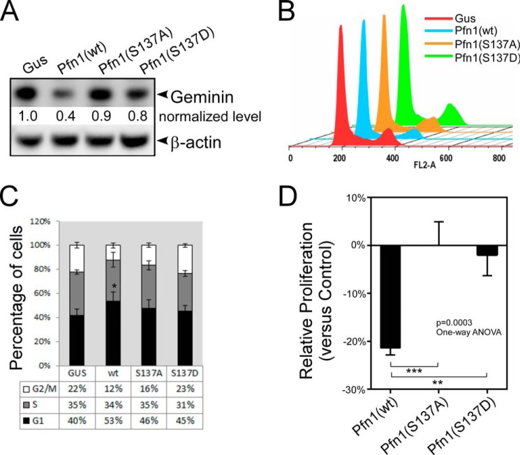
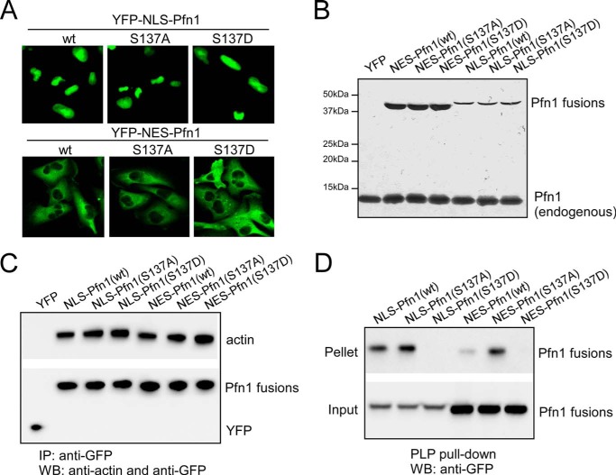
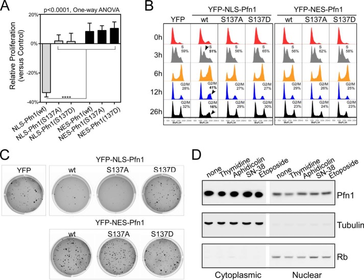
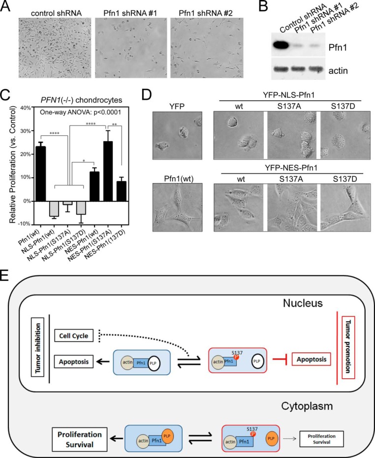
The actin-binding protein profilin-1 (Pfn1) inhibits tumor growth and yet is also required for cell proliferation and survival, an apparent paradox. We previously identified Ser-137 of Pfn1 as a phosphorylation site within the poly-l-proline (PLP) binding pocket. Here we confirm that Ser-137 phosphorylation disrupts Pfn1 binding to its PLP-containing ligands with little effect on actin binding. We find in mouse xenografts of breast cancer cells that mimicking Ser-137 phosphorylation abolishes cell cycle arrest and apoptotic sensitization by Pfn1 and confers a growth advantage to tumors. This indicates a previously unrecognized role of PLP binding in Pfn1 antitumor effects. Spatial restriction of Pfn1 to the nucleus or cytoplasm indicates that inhibition of tumor cell growth by Pfn1 requires its nuclear localization, and this activity is abolished by a phosphomimetic mutation on Ser-137. In contrast, cytoplasmic Pfn1 lacks inhibitory effects on tumor cell growth but rescues morphological and proliferative defects of PFN1 null mouse chondrocytes. These results help reconcile seemingly opposed cellular effects of Pfn1, provide new insights into the antitumor mechanism of Pfn1, and implicate Ser-137 phosphorylation as a potential therapeutic target for breast cancer.

Keywords: Apoptosis; Cancer; Cell Compartmentalization; Nucleus; Phosphorylation; Profilin; Proliferation.


Figures

FIGURE 1.
FIGURE 1.
Mimicking Ser-137 phosphorylation selectively inhibits PLP binding of Pfn1. A, x-ray diffraction structure of human Pfn1 (middle, cyan) bound with actin (left, purple) and a 16-amino acid PLP peptide derived from the vasodilator-stimulated phosphoprotein (right, green, in space-filling mode) (Protein Data Bank ID 2PAV). Ser-137 of Pfn1, indicated by the arrow, is located next to the PLP peptide but far from actin. B, HEK293 cells were transfected with HA-tagged Pfn1 (wt, S137A, or S137D), and lysates were bound to control (C) or PLP-conjugated (P) Sepharose beads. Anti-HA antibody was used to detect HA-Pfn1 bound to the beads (pellet) and in the lysate (input) by Western blot. C, HEK293 cells transfected with pcDNA3 (vector) or Myc-tagged Pfn1 (wt, S137A, and S137D) were fractionated and analyzed for F-actin (F)/G-actin (G) ratio by Western blot. Jasplakinolide was used to stabilize F-actin before fractionation as a control. Similar expression of all three Myc-Pfn1 proteins was confirmed by Western blotting using a Myc-tag antibody. D, NIH 3T3 cells were transfected with Pfn1 constructs (wt, S137A, S137D, and Y59A) and subsequently co-stained for overexpressed Pfn1 with an anti-Pfn1 antibody and F-actin with rhodamine-phalloidin. Arrowheads point to positive cells which expressed the indicated Pfn1 plasmids.
FIGURE 2.
FIGURE 2.
Mimicking Pfn1 phosphorylation on Ser-137 promotes tumor growth in vivo. A, Western blot showing stable overexpression of exogenous Pfn1 (wt, S137A, and S137D) over endogenous Pfn1 in MDA-MB-231 cells. gus, a bacterial reporter gene encoding β-glucuronidase as the negative control. β-Actin was blotted as the loading control. B–E, stable MDA-MB-231 cells were injected into the mammary fat pads of 5-week-old female NOD/SCID mice (106 cells per side, 5 mice per group). Tumor cells were monitored 1 day after injection and weekly thereafter using bioluminescence imaging (BLI). Caliper measurements started 4 weeks after injection. B, representative bioluminescence imaging images showing tumor burden at 4 weeks post-injection. C, time course of tumor growth measured by bioluminescence imaging. Total photon flux (photons/s) of 10 s/binning 2 imaging acquisition is shown for each data point. Data are the mean ± S.E. of 10 ROI (region of interest covering each entire tumor) values for each group (2 tumors per mouse, 5 mice per group). D, time course of tumor volume increase measured by caliper. Data are the mean ± S.E. of 10 tumors within each group (2 tumors per mouse, 5 mice per group). p values were based on two-tailed unpaired t test (Pfn1 versus Gus tumors). E, end-point tumor weights at resection. p values were based on one-way ANOVA and multiple comparisons using the Tukey method. *, p < 0.05; **, p < 0.01; ***, p < 0.001, ****, p < 0.0001.
FIGURE 3.
FIGURE 3.
Mimicking Pfn1 phosphorylation on Ser-137 inhibits tumor cell apoptosis in vivo. A and B, frozen tumors from the xenografted mice were analyzed for c-PARP and c-casp-3 by Western blot. Protein bands were quantified based on densitometry and normalized against β-actin levels. For simplicity, c-PARP and c-Casp-3 levels within the Gus tumor were arbitrarily set at 1, and those in Pfn1-expressing tumors were calculated accordingly. Tumors from two mice within each group were analyzed (which showed similar results), one of which is shown here. C, immunohistochemical (IHC) analysis of formalin-fixed and paraffin-embedded tumors for c-Casp-3. Nuclei were counterstained with hematoxylin. Shown are representative images at different magnifications. D, stable MDA-MB-231 cells grown in two-dimensional cultures in the absence or presence of 50 μm etoposide (24 h) were analyzed for c-PARP and c-casp-3 levels by Western blot. Etoposide was used as a positive experimental control to induce apoptosis. β-Actin was blotted as the loading control.
FIGURE 4.
FIGURE 4.
Preventing or mimicking Ser-137 phosphorylation both inhibit cell cycle arrest by Pfn1. A, frozen xenograft tumors were analyzed for geminin by Western blot. Protein bands were quantified based on densitometry and normalized against β-actin levels. Relative geminin levels in the Pfn1-expressing tumors over the Gus control tumors were shown. B and C, stable MDA-MB-231 cells expressing Gus or Pfn1 (wt, S137A, or S137D) grown in two-dimensional cultures were serum-starved for 48 h and subsequently released for 24 h. They were labeled with propidium iodide and analyzed for DNA content using flow cytometry. A representative experiment was shown in B. Data in C are the mean ± S.E. of four independent experiments. D, stable MDA-MB-231 cells expressing Gus or Pfn1 (wt, S137A, or S137D) were seeded on plastic and grown for 7 days. They were quantified by MTT labeling on day 1 and day 7, and the day 7/day 1 ratios of Pfn1-expressing cells were normalized against that of Gus control cells to give rise to their relative proliferation rates (y axis: positive % values for increase and negative % values for decrease). Data are the mean ± S.E. of five independent experiments. p values for C and D were based on one-way ANOVA analysis and multiple comparisons using the Tukey method. **, p < 0.01; ***, p < 0.001.
FIGURE 5.
FIGURE 5.
Pfn1 fused to NLS or NES maintains actin and PLP binding abilities. A, subcellular localization of YFP-NLS-Pfn1 and YFP-NES-Pfn1 in stable MDA-MB-231 cells. B, relative expression levels of YFP-NLS-Pfn1 and YFP-NES-Pfn1 in stable MDA-MB-231 cells by Western blot. C, MDA-MB-231 stable cell lysates expressing YFP, NLS-Pfn1, or NES-Pfn1 lacking or containing S137 mutations were immunoprecipitated (IP) with an anti-GFP antibody and analyzed for actin binding by Western blot (WB). D, same cell lysates from C were mixed with PLP-conjugated beads and analyzed for the binding of NLS or NES-tagged Pfn1 by Western blotting using a GFP antibody.
FIGURE 6.
FIGURE 6.
Cell cycle arrest and growth inhibition by Pfn1 requires nuclear localization. A, stable MDA-MB-231 cells were seeded in two-dimensional cultures and quantified by MTT assay at day 1 and day 7. Relative growth rates of Pfn1 fusion-expressing cells over those expressing YFP control were calculated as described in Fig. 4D. Data are the mean ± S.E. of three separate experiments. p values were based on one-way ANOVA analysis and multiple comparisons using the Tukey method. ****, p < 0.0001. B, DNA content analysis of stable MDA-MB-231 cells blocked by double thymidine and released for various lengths of time (3, 6, 12, and 26 h). Arrowheads indicated obvious changes in cell cycle profiles caused by NLS-Pfn1(wt) expression, which have been confirmed by three independent experiments. C, stable MDA-MB-231 cells were grown in 0.4% soft agar for 3 weeks and stained with crystal violet. Shown are representative images from one of three independent experiments, which generated data of similar trends. D, parental MDA-MB-231 cells were subjected to double thymidine (2 mm) block or treated with aphidicolin (5 μg/ml), SN-38 (200 nm), and etoposide (50 μm) for 24 h. Cells were fractionated, and Pfn1 in the cytoplasmic and nuclear fractions was analyzed by Western blot. Tubulin and Rb were used as cytoplasmic and nuclear markers.
FIGURE 7.
FIGURE 7.
Cytoplasmic Pfn1 rescues cellular defects of PFN1 null chondrocytes. A, equal numbers of MDA-MB-231 cells were infected with lentiviral shRNAs containing a scrambled sequence (control) or two different Pfn1-targeting sequences (#1 and #2). Cell images were taken at day 7 post-infection. B, Western blot analysis of shRNA infected MDA-MB-231 cells for total Pfn1. Actin was blotted as the loading control. C, Pfn1−/− chondrocytes infected with YFP, YFP-NLS-Pfn1, or YFP-NES-Pfn1 viruses were seeded in two-dimensional cultures and quantified by MTT assay at day 1 and day 4. Relative growth rates of Pfn1-infected cells over YFP-infected control cells were calculated as described in Fig. 5C. Data are the mean ± S.E. of three separate experiments. p values were based on one-way ANOVA analysis and multiple comparisons using the Tukey's method. *, p < 0.05; **, p < 0.01; ****, p < 0.0001. D, morphological differences of virus infected Pfn1−/− chondrocytes grown on plastic. E, A Spatial Confinement model to explain Pfn1's opposing cellular functions and the role of Ser-137 phosphorylation. Pfn1 acts as a tumor suppressor in the nucleus by augmenting apoptosis and blocking cell cycle, whereas it acts in the cytoplasm to support proliferation and survival by facilitating actin polymerization and remodeling. In the nucleus, the pro-apoptotic activity of Pfn1 depends on interaction with actin and an unknown PLP protein. Ser-137 phosphorylation blocks PLP binding, and converts Pfn1 from an apoptotic enhancer to an inhibitor. As for cell cycle inhibition, dynamic association/dissociation of Pfn1 with a different PLP protein might be required (dashed line). In the cytoplasm, Pfn1 interacts with actin and multiple PLP-containing proteins (most are actin-regulatory proteins such as mDia, Ena/VASP, and N-WASP) to support proliferation and survival. Ser-137 phosphorylation inhibits these activities by disrupting Pfn1 binding to PLP.

References

    1. Birbach A. (2008) Profilin, a multi-modal regulator of neuronal plasticity. Bioessays 30, 994–1002 - PubMed
    1. Jockusch B. M., Murk K., Rothkegel M. (2007) The profile of profilins. Rev. Physiol. Biochem. Pharmacol. 159, 131–149 - PubMed
    1. Schlüter K., Jockusch B. M., Rothkegel M. (1997) Profilins as regulators of actin dynamics. Biochim. Biophys. Acta 1359, 97–109 - PubMed
    1. Witke W. (2004) The role of profilin complexes in cell motility and other cellular processes. Trends Cell Biol. 14, 461–469 - PubMed
    1. Böttcher R. T., Wiesner S., Braun A., Wimmer R., Berna A., Elad N., Medalia O., Pfeifer A., Aszódi A., Costell M., Fässler R. (2009) Profilin 1 is required for abscission during late cytokinesis of chondrocytes. EMBO J. 28, 1157–1169 - PMC - PubMed

Publication types