. 2015 Apr 17;290(16):10395-405.
doi: 10.1074/jbc.M114.631689. Epub 2015 Feb 13.

Ubiquitin-associated domain-containing ubiquitin regulatory X (UBX) protein UBXN1 is a negative regulator of nuclear factor κB (NF-κB) signaling

Affiliations

Ubiquitin-associated domain-containing ubiquitin regulatory X (UBX) protein UBXN1 is a negative regulator of nuclear factor κB (NF-κB) signaling

Yu-Bo Wang et al. J Biol Chem. .

Abstract

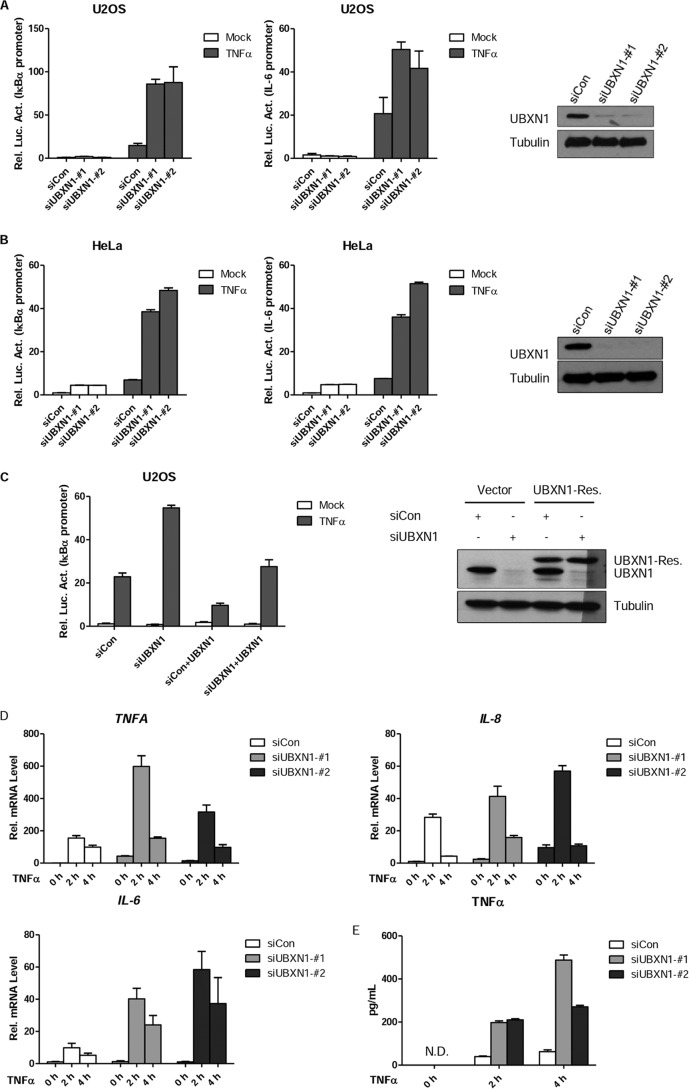
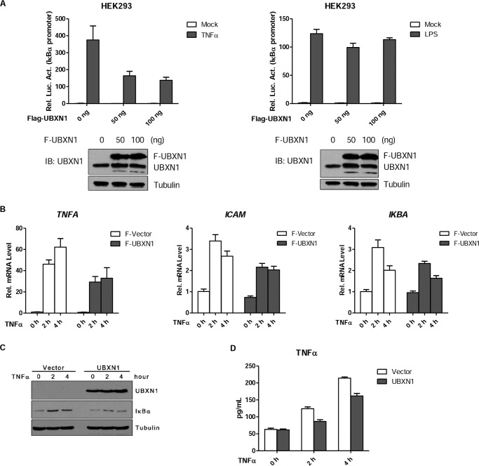
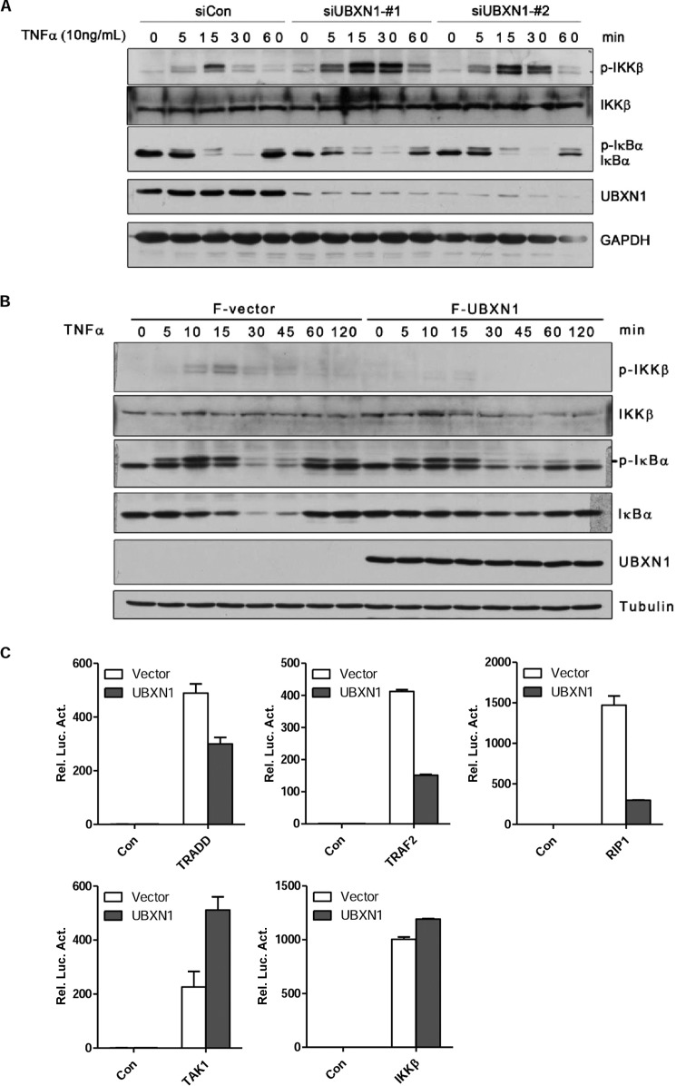
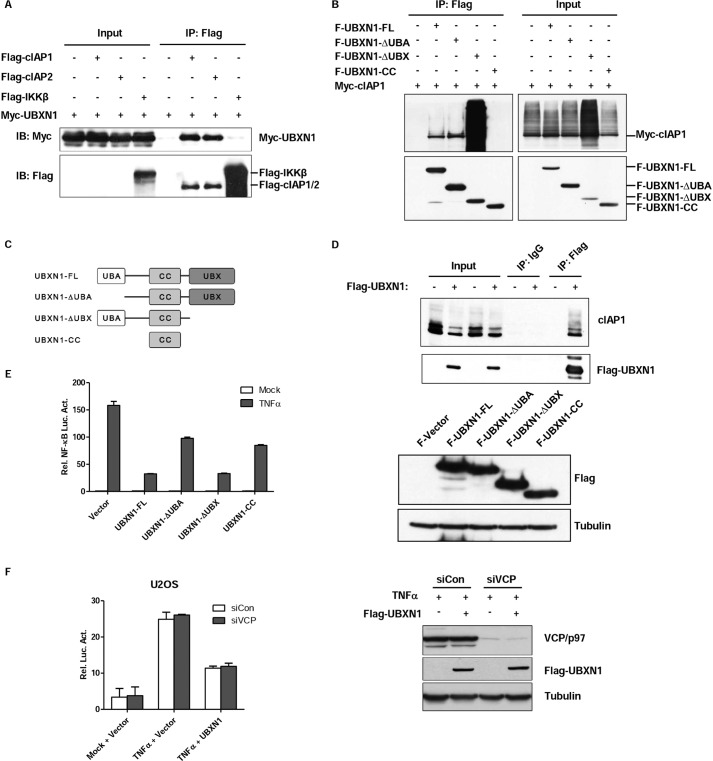
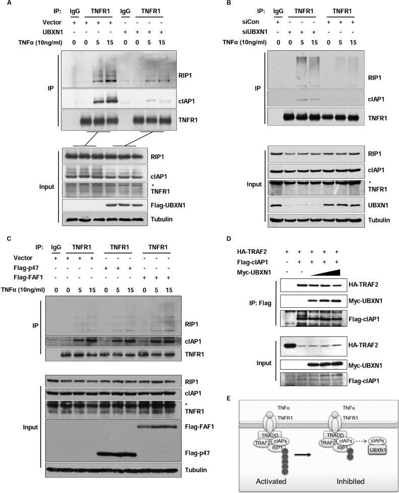
Excessive nuclear factor κB (NF-κB) activation should be precisely controlled as it contributes to multiple immune and inflammatory diseases. However, the negative regulatory mechanisms of NF-κB activation still need to be elucidated. Various types of polyubiquitin chains have proved to be involved in the process of NF-κB activation. Many negative regulators linked to ubiquitination, such as A20 and CYLD, inhibit IκB kinase activation in the NF-κB signaling pathway. To find new NF-κB signaling regulators linked to ubiquitination, we used a small scale siRNA library against 51 ubiquitin-associated domain-containing proteins and screened out UBXN1, which contained both ubiquitin-associated and ubiquitin regulatory X (UBX) domains as a negative regulator of TNFα-triggered NF-κB activation. Overexpression of UBXN1 inhibited TNFα-triggered NF-κB activation, although knockdown of UBXN1 had the opposite effect. UBX domain-containing proteins usually act as valosin-containing protein (VCP)/p97 cofactors. However, knockdown of VCP/p97 barely affected UBXN1-mediated NF-κB inhibition. At the same time, we found that UBXN1 interacted with cellular inhibitors of apoptosis proteins (cIAPs), E3 ubiquitin ligases of RIP1 in the TNFα receptor complex. UBXN1 competitively bound to cIAP1, blocked cIAP1 recruitment to TNFR1, and sequentially inhibited RIP1 polyubiquitination in response to TNFα. Therefore, our findings demonstrate that UBXN1 is an important negative regulator of the TNFα-triggered NF-κB signaling pathway by mediating cIAP recruitment independent of VCP/p97.

Keywords: Inflammation; NF-κB; Polyubiquitin Chain; Signal Transduction; Tumor Necrosis Factor (TNF); UBXN1; cIAPs.


Figures

FIGURE 1.
FIGURE 1.
RNAi screen of UBA domain proteins that regulate NF-κB activity. A–C, small scale RNAi screen using a library targeting UBA domain proteins screened out UBXN1 as a potential NF-κB negative regulator. A, screening with siRNAs against 51 known/predicted UBA domain proteins was performed using the Dharmacon SMARTpool® siRNA library, in which each siRNA consisted of four individual sequences. HeLa cells were transfected with NF-κB luciferase plasmid and siRNAs. 48 h after transfection, the cells were treated with TNFα (10 ng/ml) or left untreated (NT) for 10 h before luciferase assays were performed. The gene symbols in the graph's horizontal axis represent related siRNA-targeted genes. The average raw luciferase value of the screen was inferred and indicated (Aver.). Values that are 1.2-fold higher or 1.2-fold lower than the average were marked by dashed lines. B, dual-luciferase assays of NF-κB activity in TNFα-treated HeLa cells transfected with control siRNA or siRNA against UBA-UBX family members, including UBXN1, NSFL1C, UBXD7, UBXD8, and FAF1. C, dual-luciferase assays of NF-κB activity in TNFα-treated HEK293 cells overexpressing control vector or UBA-UBX family members, including UBXN1, NSFL1C, UBXD7, UBXD8, and FAF1.
FIGURE 2.
FIGURE 2.
Knockdown of UBXN1 potentiates TNFα-triggered NF-κB signaling. A, B, effects of UBXN1 knockdown on TNFα-triggered NF-κB activation in U2OS cells and HeLa cells. U2OS cells or HeLa cells (1 × 10⁵) were first transfected with the pNF-κB-Luc (IκBα promoter, left), IL-6-Luc (IL-6 promoter, middle), and pRL-TK plasmids, and then control siRNA or siRNA against UBXN1-#1 and UBXN1-#2 12 h later. 48 h after siRNA transfection, cells were treated with TNFα (10 ng/ml) or left untreated for 10 h before luciferase assays were performed. Knockdown efficiencies of indicated siRNAs in U2OS cells were examined by immunoblot analysis (right). C, effects of UBXN1 knockdown on TNFα-triggered NF-κB activation could be rescued by UBXN1 expression. U2OS cells (2 × 10⁵) were transfected with control siRNA or siRNAs against UBXN1-#1, 24 h later, UBXN1-rescue plasmid was transfected for another 24 h. Cells were treated with TNFα (10 ng/ml) or left untreated for 10 h before luciferase assays were performed. The experiments were similarly performed as in A. D, effects of UBXN1 knockdown on TNFα-induced transcription of TNFA, IL-8, and IL-6 genes. U2OS cells (2 × 10⁵) were transfected with control siRNA or siRNAs against UBXN1-#1 and UBXN1-#2. Forty eight hours later, cells were treated with TNFα for the indicated times, and then total RNA was prepared for qPCR analysis. Expression is presented relative to GAPDH expression. E, effects of UBXN1 knockdown on TNFα-induced cytokine production of TNFα. U2OS cells (4 × 10⁵) were transfected with control siRNA or siRNAs against UBXN1-#1 and UBXN1-#2. Forty eight hours later, cells were treated with TNFα for the indicated times, and then total supernatant was prepared for ELISA. N.D., not detected.
FIGURE 3.
FIGURE 3.
Overexpression of UBXN1 inhibits TNFα-triggered NF-κB activation. A, effects of UBXN1 overexpression on TNFα-triggered NF-κB activation in HEK293 cells were examined by dual-luciferase assays. HEK293 cells (1 × 10⁵) were transfected with the pNF-κB-Luc, pRL-TK, and increasing amounts of FLAG-UBXN1 plasmid. 48 h after transfection, cells were treated with TNFα (10 ng/ml), LPS (1 μg/ml), or left untreated for 10 h before luciferase assays were performed. Expression of UBXN1 in HEK293 cells were examined by immunoblot analysis (bottom). B, effects of UBXN1 overexpression on TNFα-induced transcription of TNFA, ICAM, and IKBA genes. HEK293 (2 × 10⁵) transfected with either an empty vector or FLAG-UBXN1 plasmid were treated with TNFα (10 ng/ml) for the indicated times, and then total RNA was prepared for qPCR analysis. The mRNA expression is presented relative to GAPDH expression. C, protein expression of UBXN1 and IκBα at indicated times after TNFα (10 ng/ml) treatment was examined by immunoblot analysis. D, effects of UBXN1 overexpression on TNFα-induced cytokine production of TNFα. HeLa cells (4 × 10⁵) were transfected with either an empty vector or FLAG-UBXN1 plasmid. 36 h later, cells were treated with TNFα for the indicated times, and then total supernatant was prepared for ELISA.
FIGURE 4.
FIGURE 4.
UBXN1 inhibits TNFα-triggered NF-κB signaling at TNF receptor level. A and B, UBXN1 inhibits the process of TNFα-triggered NF-κB activation. A, U2OS cells (2 × 10⁵) were transfected with control siRNA, UBXN1-#1, and UBXN1-#2. After 72 h, the cells were treated with TNFα (10 ng/ml) for indicated times. The whole cell lysate was analyzed by immunoblotting with the indicated antibodies. B, HEK293T cells (2 × 10⁵) were transfected with either an empty vector or FLAG-UBXN1 plasmid. After 36 h, cells were treated with TNFα (10 ng/ml) for the indicated times. The whole cell lysate was analyzed by immunoblotting with the indicated antibodies. C, UBXN1 inhibits TRADD-, TRAF2-, and RIP1-mediated but not TAK1- or IKKβ-mediated NF-κB activation. HEK293T cells (1 × 10⁵) were transfected with reporter plasmid, either an empty vector, or FLAG-UBXN1 plasmid together with TRADD, TRAF2, RIP1, TAK1, IKKβ, respectively. Luciferase assay was assessed 24 h after transfection.
FIGURE 5.
FIGURE 5.
UBA domain of UBXN1 is crucial for UBXN1-mediated NF-κB inhibition. A–D, UBXN1 interacts with cIAPs. A, immunoassay of lysates of HEK293T cells cotransfected with vectors expressing FLAG-tagged (Flag-) cIAP1, cIAP2, or IKKβ and Myc-tagged (Myc-) UBXN1, immunoprecipitated (IP) with anti-FLAG, and analyzed by immunoblotting (IB) with anti-Myc. B, UBXN1 interacts with cIAP1 through either the UBA domain or UBX domain. Coimmunoprecipitation of cIAP1 and UBXN1 from HEK293T cells expressing Myc-cIAP1 and FLAG-tagged UBXN1 mutants using anti-FLAG-agarose beads was followed by immunoblot using a FLAG or Myc antibody. C, schematic representation of UBXN1 deletion mutants. D, immunoblot analysis of the interaction between endogenous cIAP1 proteins and exogenous UBXN1 in lysates of HEK293T cells transfected with either an empty vector or FLAG-UBXN1 plasmid after immunoprecipitation with mouse IgG or anti-FLAG (right) or with the same amount of cell lysate used for immunoprecipitation (Input; left). E, UBA domain of UBXN1 is essential for inhibiting TNFα-triggered NF-κB activation. Dual-luciferase assays of NF-κB activity in TNFα-treated HEK293T cells overexpressing FLAG-tagged UBXN1 truncates as described in C. The expression levels of FLAG-UBXN1 truncated mutants were examined by immunoblots. F, knockdown of VCP/p97 does not have effect on UBXN1-mediated NF-κB inhibition. The experiment was similarly performed as in Fig. 2A.
FIGURE 6.
FIGURE 6.
UBXN1 sequesters cIAPs from TRAF2 and decreases RIP1 ubiquitination. A, overexpression of UBXN1 inhibits cIAP1 recruitment to TNFR1 and RIP1 ubiquitination upon TNFα stimulation. HEK293T cells transfected with vectors or FLAG-tagged (Flag-) UBXN1 plasmids were left untreated or treated with TNFα (10 ng/ml) for the indicated times. Immunoprecipitation (IP) and immunoblot analysis were performed with the indicated antibodies. B, UBXN1 deficiency potentiates cIAP1 recruitment to TNFR1 and RIP1 ubiquitination upon TNFα stimulation. U2OS cells transfected with control siRNA or siRNA against UBXN1 were left untreated or treated with TNFα (10 ng/ml) for the indicated times. Immunoprecipitation and immunoblot analysis were performed with the indicated antibodies. C, overexpression of NSFL1C/p47 and FAF1 barely inhibit cIAP1 recruitment to TNFR1 and RIP1 ubiquitination upon TNFα stimulation. Similar experiments were performed as in A. D, immunoassay of HEK293T cells transfected with HA-tagged TRAF2, FLAG-tagged cIAP1, and increasing amounts (wedges) of Myc-tagged UBXN1; cIAP1 in lysates was immunoprecipitated with anti-FLAG, followed by immunoblot analysis of immunoprecipitates (top) and input lysate (bottom). E, model illustrating UBXN1-mediated TNFα-triggered NF-κB signaling inhibition. Upon TNF-α stimulation, TRAF2 and RIP1 were recuited to TNFR1. cIAPs associate with TRAF2 and catalyze the conjugation of Lys-63-linked polyubiquitin chains to RIP1. UBXN1 binds to cIAPs and sequesters cIAPs from TRAF2 and decreases RIP1 ubiquitination.

Similar articles

Cited by

References

    1. Karin M. (2006) Nuclear factor-κB in cancer development and progression. Nature 441, 431–436 - PubMed
    1. Hayden M. S., Ghosh S. (2012) NF-κB, the first quarter-century: remarkable progress and outstanding questions. Genes Dev. 26, 203–234 - PMC - PubMed
    1. DiDonato J. A., Mercurio F., Karin M. (2012) NF-κB and the link between inflammation and cancer. Immunol. Rev. 246, 379–400 - PubMed
    1. Chen G., Goeddel D. V. (2002) TNF-R1 signaling: a beautiful pathway. Science 296, 1634–1635 - PubMed
    1. Wajant H., Pfizenmaier K., Scheurich P. (2003) Tumor necrosis factor signaling. Cell Death Differ. 10, 45–65 - PubMed

Publication types

MeSH terms

LinkOut - more resources