Case Reports
. 2015 Mar 5;96(3):432-9.
doi: 10.1016/j.ajhg.2015.01.002. Epub 2015 Feb 12.

Mutations in SEC24D, encoding a component of the COPII machinery, cause a syndromic form of osteogenesis imperfecta

Affiliations
Case Reports

Mutations in SEC24D, encoding a component of the COPII machinery, cause a syndromic form of osteogenesis imperfecta

Lutz Garbes et al. Am J Hum Genet. .

Abstract

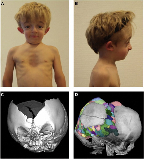
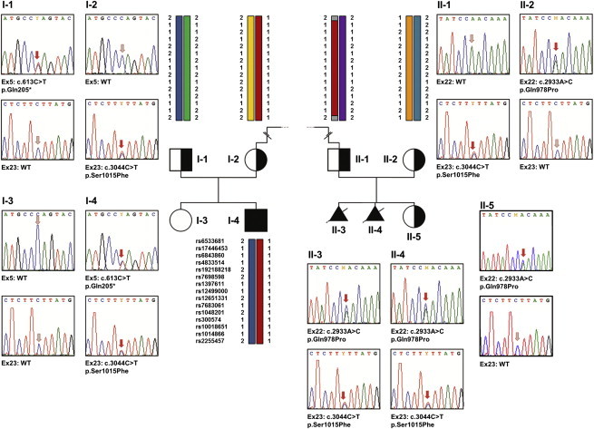
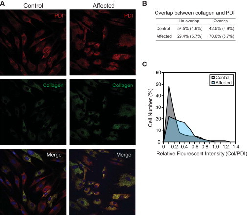
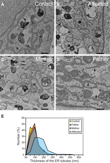
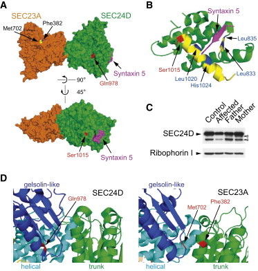
As a result of a whole-exome sequencing study, we report three mutant alleles in SEC24D, a gene encoding a component of the COPII complex involved in protein export from the ER: the truncating mutation c.613C>T (p.Gln205(∗)) and the missense mutations c.3044C>T (p.Ser1015Phe, located in a cargo-binding pocket) and c.2933A>C (p.Gln978Pro, located in the gelsolin-like domain). Three individuals from two families affected by a similar skeletal phenotype were each compound heterozygous for two of these mutant alleles, with c.3044C>T being embedded in a 14 Mb founder haplotype shared by all three. The affected individuals were a 7-year-old boy with a phenotype most closely resembling Cole-Carpenter syndrome and two fetuses initially suspected to have a severe type of osteogenesis imperfecta. All three displayed a severely disturbed ossification of the skull and multiple fractures with prenatal onset. The 7-year-old boy had short stature and craniofacial malformations including macrocephaly, midface hypoplasia, micrognathia, frontal bossing, and down-slanting palpebral fissures. Electron and immunofluorescence microscopy of skin fibroblasts of this individual revealed that ER export of procollagen was inefficient and that ER tubules were dilated, faithfully reproducing the cellular phenotype of individuals with cranio-lentico-sutural dysplasia (CLSD). CLSD is caused by SEC23A mutations and displays a largely overlapping craniofacial phenotype, but it is not characterized by generalized bone fragility and presented with cataracts in the original family described. The cellular and morphological phenotypes we report are in concordance with the phenotypes described for the Sec24d-deficient fish mutants vbi (medaka) and bulldog (zebrafish).


Figures

Figure 1
Figure 1
Photographs and CT Scans of the Affected Individual from Family 1 (A) Affected individual from family 1 at the age of 7 years. Note facial dysmorphism with down-slanting palpebral fissures, hypertelorism, dysplastic right ear, and pectus excavatus. (B) Lateral view. Note mid-face hypoplasia, micrognathia, frontal bossing, and dysplastic right ear. (C) Three-dimensional CT scan of the cranium at the age of 14 months. Note wide sagittal suture with a broad ossification defect fronto-parietal apical. Sutura metopica is displaced to the bottom, as indicated by surrounding wormian bones. Coronal sutures, lambda sutures, and temporal sutures appear narrow and with premature ossification pattern without clear synostosis, but with multiple wormian bones enclosed. (D) Lateral view of a cranial CT scan taken at age 4 years and 4 months, with multiple wormian bones highlighted in different colors. Note an intraparietal suture on the left side (described as unilateral intraparietal suture30). The large ossification defect fronto-parietal apical persisted. The sphenoid wings appear with an increased density and with multiple erosions.
Figure 2
Figure 2
Validation of the SEC24D Mutations by Sanger Sequencing of Genomic DNA Pedigrees of the two analyzed families and electropherograms of the respective SEC24D mutations in exons 5, 22, and 23 are shown. In family 1, each parent carries one of the two mutations and the affected son (I-4) is compound heterozygous for the SEC24D mutations c.613C>T (p.Gln205) and c.3044C>T (p.Ser1015Phe). The healthy sister of the affected individual did not carry either of the two mutations. Also in family 2, each parent is heterozygous carrier of one of the two SEC24D mutations and both affected fetuses (II-3 and II-4) are compound heterozygous for c.2933A>C (p.Gln978Pro) and c.3044C>T (p.Ser1015Phe). The healthy sister (II-5) is a heterozygous carrier of only one of these mutations. The red arrows in the electropherograms indicate either the mutation or the respective position in the wild-type sequence. In addition, for several family members the haplotype combination at the SEC24D locus is shown, and 15 of the 166 consecutive good-quality SNPs used for reconstructing the 14 Mb disease haplotype are depicted. Note that individuals I-2, I-4, and II-1 share the SEC24D mutation c.3044C>T and an identical haplotype between the SNPs rs6533681 and rs2255457. The dashed line between I-2 and II-2 indicates that the mutation c.3044C>T is a founder mutation inherited from a distant common ancestor.
Figure 3
Figure 3
Retention of Procollagen in the ER (A) Double immunofluorescent labeling of control and the affected individual’s fibroblasts for type I procollagen and protein disulfide isomerase (PDI), an ER marker. Images were obtained by standard confocal microscopy. (B) The number of cells with an indicated overlap of procollagen with PDI was shown. Standard deviations from three independent experiments are shown in parentheses. (C) ImageJ was used to normalize fluorescent intensities of procollagen with those of PDI from the images obtained via confocal microscopy. Procollagen is retained more in the fibroblasts of the affected individual (light blue) than in the control fibroblasts (gray) with statistical significance (Student’s t test, p = 0.0001, n = 467 cells for control fibroblast, n = 554 cells for the affected individual’s fibroblasts).
Figure 4
Figure 4
Dilation of ER in the Fibroblasts of the Affected Individual from Family 1 (A–D) Micrographs of thin-section electron microscopy for fibroblasts from control (A), affected individual (B), mother of affected individual (C), and father of affected individual (D). Tubular ER elements studded with ribosomes are indicated by arrowheads. Scale bars represent 500 nm. (E) Summary of the thickness of ER tubules in fibroblasts. 100 nm in the x axis represents a thickness that is larger than 50 nm but equal to or less than 100 nm. The average thickness of the tubules is about 189 nm, 120 nm, 116 nm, and 125 nm for the cells from the affected individual (light blue), control (yellow), father (orange), and mother (gray), respectively. The ER tubules of the fibroblasts from the affected individual were thicker than those of other fibroblasts with statistical significance according to a one-way ANOVA with Tukey’s honest significant difference test (p < 0.01; n = 183 tubules for control, 212 for father, 171 for mother, and 181 for the affected individual).
Figure 5
Figure 5
Position of the Mutant Residues in the 3D Structure of SEC24D Note that the available human SEC24D structures were solved with the isoform 2 (O94855-2) (1,033 aa). Here we used the nomenclature of the isoform 1 (O94855-1) (1,032 aa) of SEC24D because this form has been chosen as the canonical sequence in the databases. PDB 3EFO structure was used. (A) Gln978 (red, upper panel) is located in the gelsolin-like domain of SEC24D. The location of Gln978 SEC24D is structurally similar to that of Met702 (yellow) and Phe382 (blue) of SEC23A. A more detailed structural comparison was shown in (D). Ser1015 (red, lower panel) is found in the IxM pocket (I, x, and M stand for Ile, any amino acid, and Met). A fragment of syntaxin 5 (magenta) contains an IxM signal. A more detailed structure is shown in (B). (B) The helical domain of SEC24D is shown. Ser1015 (red) is located in an α-helix (yellow) that contains key residues (Leu1020 and His1024, blue) of the IxM pocket. Additional key residues (Leu833 and Leu835, blue) of this pocket are located in a β-strand (yellow). (C) Total lysates (10 μg) of indicated fibroblasts derived from family 1 were prepared, resolved by SDS-PAGE, and processed for immunoblotting. Control foreskin fibroblasts were obtained from the American Type Culture Collection (ATCC, CRL-091). Ribophorin 1 (an ER-resident protein) was probed as loading controls. Bands (open arrowheads) below the main SEC24D band probably represent partial degradation products of SEC24D or nonspecific proteins. (D) The gelsolin-like, the helical, and the trunk domains of SEC24D (left) and SEC23A (right) were colored blue, cyan, and green, respectively. CLSD-linked mutations were colored red. Met702 and Phe382 of SEC23A constitute a part of the SEC31-binding pocket.

References

    1. Rauch F., Glorieux F.H. Osteogenesis imperfecta. Lancet. 2004;363:1377–1385. - PubMed
    1. Canty E.G., Kadler K.E. Procollagen trafficking, processing and fibrillogenesis. J. Cell Sci. 2005;118:1341–1353. - PubMed
    1. Forlino A., Cabral W.A., Barnes A.M., Marini J.C. New perspectives on osteogenesis imperfecta. Nat. Rev. Endocrinol. 2011;7:540–557. - PMC - PubMed
    1. Byers P.H., Pyott S.M. Recessively inherited forms of osteogenesis imperfecta. Annu. Rev. Genet. 2012;46:475–497. - PubMed
    1. Marini J.C., Reich A., Smith S.M. Osteogenesis imperfecta due to mutations in non-collagenous genes: lessons in the biology of bone formation. Curr. Opin. Pediatr. 2014;26:500–507. - PMC - PubMed

Publication types

MeSH terms

Substances

Supplementary concepts