Review
. 2015 May:479-480:545-54.
doi: 10.1016/j.virol.2015.01.014. Epub 2015 Feb 13.

Initiation and regulation of paramyxovirus transcription and replication

Affiliations
Review

Initiation and regulation of paramyxovirus transcription and replication

Sarah L Noton et al. Virology. 2015 May.

Abstract

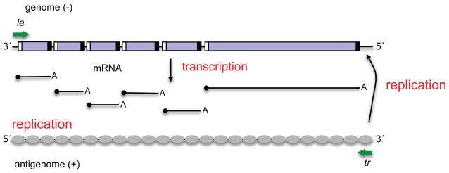
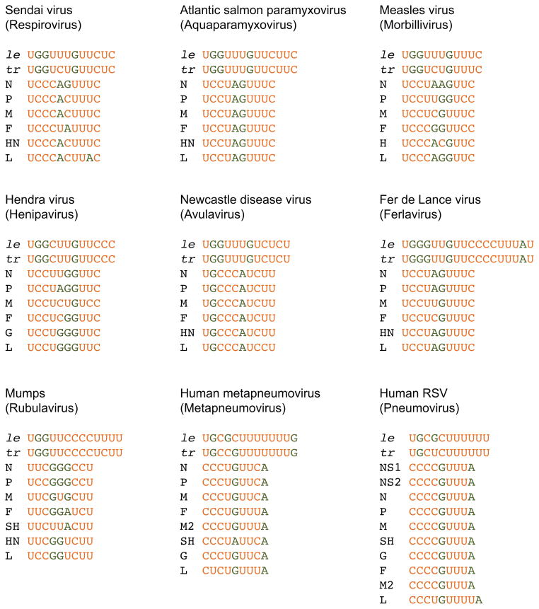
The paramyxovirus family has a genome consisting of a single strand of negative sense RNA. This genome acts as a template for two distinct processes: transcription to generate subgenomic, capped and polyadenylated mRNAs, and genome replication. These viruses only encode one polymerase. Thus, an intriguing question is, how does the viral polymerase initiate and become committed to either transcription or replication? By answering this we can begin to understand how these two processes are regulated. In this review article, we present recent findings from studies on the paramyxovirus, respiratory syncytial virus, which show how its polymerase is able to initiate transcription and replication from a single promoter. We discuss how these findings apply to other paramyxoviruses. Then, we examine how trans-acting proteins and promoter secondary structure might serve to regulate transcription and replication during different phases of the paramyxovirus replication cycle.

Keywords: Gene expression; Mononegavirales; Non-segmented negative strand RNA virus; Paramyxoviridae; Paramyxovirus; Polymerase; Promoter; Replication; Transcription.


Figures

Figure 1
Figure 1
Schematic diagram illustrating a representative paramyxovirus genome and transcription and RNA replication products. The genes are represented by purple boxes, and gs and ge signals are illustrated with white and black boxes, respectively. The le and tr promoters at the 3′ ends of the genome and antigenome, respectively, are indicated with green arrows. The genome acts as a template for mRNA and antigenome synthesis, and the antigenome as a template for genome RNA synthesis. The mRNA caps are indicated with black circles. The antigenome is shown covered with grey ovals, representing N protein, to indicate that it is encapsidated. The genome is also encapsidated, but this is not shown so that the cis-acting signals can be clearly seen.
Figure 2
Figure 2
Schematic diagrams of the le and gs regions of RSV (a paramyxovirus) and VSV (a rhabdovirus) showing the positions of the transcription-specific signals in the respective viruses. Regions in which mutations reduced transcription to less than 15% of wt levels are shown in red. Regions in which mutations reduced transcription to 15–40% of wt levels are shown in orange. Substitution of the region shown in gray had no effect on transcription. The numbers underneath indicate nucleotide positions. It should be noted that not all signals have been mapped precisely.
Figure 3
Figure 3
Diagram showing the alignment of the RSV le and L gs sequences. The figure shows the 3′ terminal 15 nt of the le and the first 10 nt of the L gene. The nucleotides shown in red in the le region were shown to be essential for both transcription and replication. These nucleotides align with the L gs signal. The green arrows show the experimentally determined initiation sites at positions 1 and 3 of le, which we propose are the replication and transcription initiation sites, respectively.
Figure 4
Figure 4
Diagrams showing the models for initiation of transcription (A) and replication (B) in RSV infection. The figure shows the relationship between the polymerase, NTPs, the le promoter region and gs signal in each case. The le region is shown as three sections, reflecting the distribution of cis-acting signals, as determined by mapping analysis. The first eleven nucleotides of the le region are written, with the core promoter element shown in red type. The black line in the middle of le indicates sequence required specifically for replication, and the white box at the end of le indicates a U-rich sequence that enhances transcription. The gs signal is shown as a large white box. In the replication initiation model (B), N protein binding to the nascent le+ RNA allows encapsidation. N is likely delivered to the RNA as a soluble N0P complex, but the P protein is not shown for simplicity.
Figure 5
Figure 5
Promoter and gs sequences for one virus species from each of the paramyxovirus genera (indicated in parentheses). The gs signals for each of the viral genes are shown. The promoter sequences shown were defined as those nucleotides that were identical or almost identical between the le and tr promoters; the minimal core promoters might be more constrained than what is shown. Pyrimidine and purine residues are shown in orange and green type, respectively.

Similar articles

Cited by

References

    1. King AMQ, Adams MJ, Carstens EB, Lefkowitz EJ. Virus taxonomy: classification and nomenclature of viruses: Ninthe Report of the International Committee on Taxonomy of Viruses. Elsevier Academic Press; 2012.
    1. Lamb RA, Parks GD. Paramyxoviridae: the viruses and their replication. In: Knipe DM, Howley PM, editors. Fields Virology. 5. Lippincott Williams and Wilkins; pp. 1449–1496.
    1. Whelan SP, Barr JN, Wertz GW. Transcription and replication of nonsegmented negative-strand RNA viruses. Curr Top Microbiol Immunol. 2004;283:61–119. - PubMed
    1. Morin B, Kranzusch PJ, Rahmeh AA, Whelan SP. The polymerase of negative-stranded RNA viruses. Current opinion in virology. 2013;3:103–110. - PMC - PubMed
    1. Horikami SM, Curran J, Kolakofsky D, Moyer SA. Complexes of Sendai virus NP-P and P-L proteins are required for defective interfering particle genome replication in vitro. J Virol. 1992;66:4901–4908. - PMC - PubMed

Publication types

MeSH terms