. 2015 Jun;145(2):300-11.
doi: 10.1111/imm.12450.

Improved emergency myelopoiesis and survival in neonatal sepsis by caspase-1/11 ablation

Affiliations

Improved emergency myelopoiesis and survival in neonatal sepsis by caspase-1/11 ablation

Lori F Gentile et al. Immunology. 2015 Jun.

Abstract

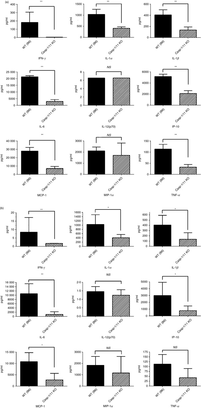
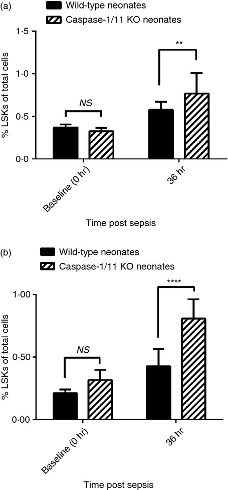
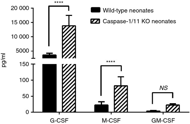
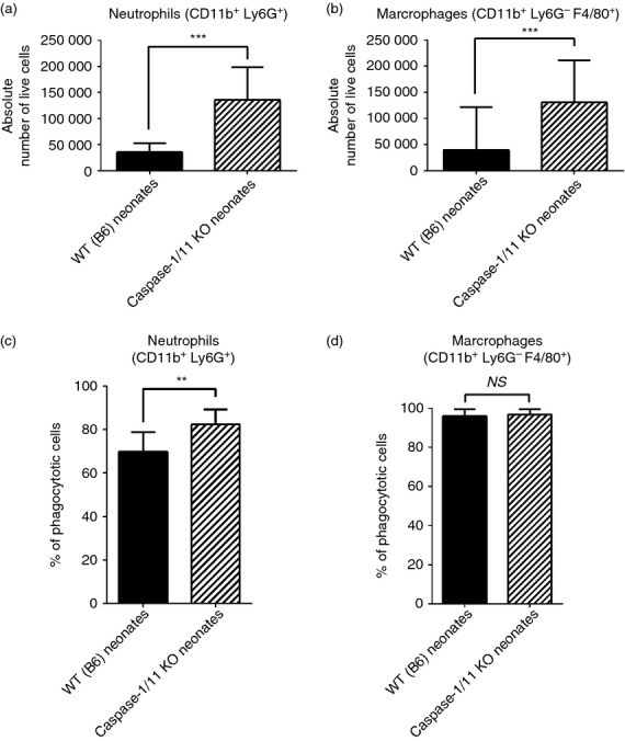
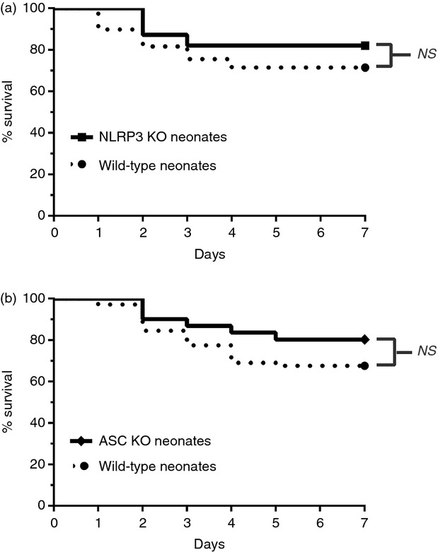
Over one million newborns die annually from sepsis with the highest mortality in premature and low-birthweight infants. The inflammasome plays a central role in the regulation of innate immunity and inflammation, and is presumed to be involved in protective immunity, in large part through the caspase-1-dependent activation of interleukin-1β (IL-1β) and IL-18. Studies in endotoxic shock, however, suggest that endogenous caspase-1 activity and the inflammasome contribute to mortality primarily by promoting excessive systemic inflammatory responses. We examined whether caspase-1 and the inflammasome also regulate neonatal inflammation, host protective immunity and myelopoiesis during polymicrobial sepsis. Neonatal (5-7 days) C57BL/6 and caspase-1/11(-/-) mice underwent a low-lethality caecal slurry model of intra-abdominal sepsis (LD25-45 ). Ablation of caspase-1/11, but not apoptosis-associated speck-like protein containing a CARD domain or nucleotide-binding oligomerization domain-like receptor protein 3 (NLRP3), improved neonatal survival following septic challenge compared with wild-type mice (P < 0·001), with decreased concentrations of inflammatory cytokines in the serum and peritoneum. Surprisingly, caspase-1/11(-/-) neonates also exhibited increased bone marrow and splenic haematopoietic stem cell expansion (P < 0·001), and increased concentrations of granulocyte and macrophage colony-stimulating factors in the peritoneum (P < 0·001) after sepsis. Ablation of caspase-1/11 signalling was also associated with increased recruitment of peritoneal macrophages and neutrophils (P < 0·001), increased phagocytosis by neutrophils (P = 0·003), and decreased bacterial colonization (P = 0·02) in the peritoneum. These findings suggest that endogenous caspase-1/11 activity, independent of the NLRP3 inflammasome, not only promotes the magnitude of the inflammatory response, but also suppresses protective immunity in the neonate, so contributing to innate immune dysfunction and poor survival in neonatal sepsis.

Keywords: caspase-1/11; emergency myelopoiesis; inflammation; mouse; neonates.


Figures

Figure 1
Figure 1
Caspase-1/11−/− neonatal mice have superior survival following intra-abdominal polymicrobial sepsis compared with wild-type (WT) mice. Caspase-1/11−/− neonates (5–7 days) and juveniles (6–8 week) and WT neonates (5–7 days) and juveniles (6–8 weeks) were challenged via intraperitoneal injection of polymicrobial caecal slurry (1·1 mg/g body weight) to achieve an LD25–45. Mice were followed for 7 days to assess survival. (a) Caspase-1/11−/− (Casp-1/11 KO) neonates (5–7 days) and juveniles (6–8 week) and WT (WT B6) neonates (5–7 days) and juveniles (6–8 weeks) were challenged with caecal slurry simultaneously. Caspase-1/11−/− neonatal mice had the greatest survival compared with juvenile caspase-1/11−/− and WT mice (neonates and juveniles) (P < 0·05, log-rank Mantel–Cox Test) (Caspase-1/11 juveniles n = 30, WT juveniles n = 30). (b) Caspase-1/11−/− (Casp-1/11 KO) neonates (5–7 days) had improved survival to caecal slurry induced sepsis compared with wild-type neonates (Caspase-1/11−/− n = 40, WT n = 42) (P < 0·001, Log Rank test).
Figure 2
Figure 2
Caspase-1/11−/− neonatal mice have decreased production of inflammatory cytokines and chemokines 18 hr after sepsis compared with wild-type (WT) mice. Neonatal (5–7 day) caspase-1/11−/− (Casp-1/11 KO) and WT (WT B6) mice were challenged with intraperitoneal polymicrobial caecal slurry (1·1 mg/g body weight; LD25–45). Serum and peritoneal washes were collected at 18 and 24 hr following sepsis and analysed by Luminex™ assay for cytokine/chemokine concentrations. (a) Caspase-1/11−/− neonatal mice (hashed bars) had decreased concentrations of interferon-γ (IFN-γ), IL-1α, IL-1β, IL-6, IFN-γ inducible protein-10 (IP-10), macrophage inflammatory protein-1 (MCP-1) and tumour necrosis factor-α (TNF-α) in the serum compared with WT neonates (solid bars). (b) Caspase-1/11−/− neonatal mice (hashed bars) have decreased concentrations of IFN-γ, IL-1α, IL-1β, IL-6, IP-10, MCP-1, and TNF-α in the peritoneum compared with WT mice (solid bars). Symbols placed within the graph show all significant differences between groups for specific cytokine. NS = non-significant, *P < 0·01, **P ≤ 0·001, Student's t-test. (n = 4 or 5 neonates per group). Data in (a) and (b) is representative of three independent experiments.
Figure 3
Figure 3
Caspase-1/11−/− mice have increased expansion of splenic and bone marrow lineage sca-1+ c-kit+ (LSK) progenitor cells in the bone marrow 36 hr after sepsis. Caspase-1/11−/− (Casp-1/11 KO) and wild-type (WT B6) neonates (5–7 days) were challenged with polymicrobial caecal slurry (1·1 mg/g body weight) via intraperitoneal injection. Bone marrow and splenocytes were collected at 36 hr and analysed by flow cytometry for the presence of lineage sca-1+ c-kit+ (LSKs). Bone marrow and splenocytes were collected from untreated neonates to establish baseline (0 hr) LSK levels. A minimum of 1 × 104 non-debris live cells were collected for analysis. Both neonatal WT and caspase-1/11−/− mice had increased splenic and bone marrow LSKs 36 hr after sepsis (P < 0·0001, two-way analysis of variance with Bonferroni multiple comparison's post hoc test). (a) Caspase-1/11−/− neonates (hashed bars) had a significantly greater expansion of bone marrow LSKs compared with WT (solid bars) neonates (57·4% versus 24·5%; **P < 0·01, Student's t-test, n = 13 or 14/group). (b) Caspase-1/11−/− neonates (hashed bars) demonstrated more than a twofold increased expansion of splenic LSKs 36 hr following sepsis compared with WT (solid bars) mice (43% versus 18%; ****P < 0·0001, Student's t-test, n = 13 or 14/group). Data shown are from three or more independent experiments. Values are expressed as per cent total of all viable cells.
Figure 4
Figure 4
Caspase-1/11−/− mice have increased concentrations of local growth factors 24 hr after sepsis. Caspase-1/11−/− (Casp-1/11 KO) and wild-type (WT B6) neonates (5–7 days) were challenged with intraperitoneal polymicrobial caecal slurry as described (LD25–45). Peritoneal washes were collected at 24 hr and analysed by Luminex™ assay for the presence of granulocyte (G-CSF), macrophage (M-CSF) and granulocyte–macrophage (GM-CSF) colony-stimulating factors. Caspase-1/11−/− (hashed bars) neonates had increased production of local growth factors (G-CSF, M-CSF) in the peritoneum 24 hr after sepsis compared with WT (solid bars) mice (****P < 0·0001, NS = no statistical significance, two-way analysis of variance with Bonferonni's multiple comparisons test) (n = 5 to n = 8 neonates/group). Data shown are from three or more independent experiments.
Figure 5
Figure 5
Caspase-1/11-deficient neonates have increased recruitment and function of innate immune effector cells after sepsis. Caspase-1/11 KO neonates (Casp-1/11 KO) and wild-type (WT B6) neonates (5–7 days) were challenged with intraperitoneal caecal slurry as described (LD25–45). Peritoneal washes were collected at 18 and 24 hr following sepsis, stained and analysed by flow cytometery to determine cell phenotype. Erythrocytes were lysed with ammonium chloride lysis buffer. (a) Caspase-1/11−/− neonates (hashed bars) had increased absolute numbers of neutrophils (CD11b+ Ly6G+) and (b) macrophages (CD11b+ Ly6G F4/80+) in the peritoneum 18 hr after sepsis compared with WT (solid bars) neonates (***P < 0·001, Student's t-test) (n = 4 or 5 neonates/group (a,b)). (c) Caspase-1/11−/− (hashed bars) had significantly increased neutrophil (CD11b+ Ly6G+) phagocytosis compared with WT (solid bars) mice (**P = 0·003, Student's t-test) (n = 5 to n = 8 neonates/group). (d) There was no difference in macrophage (CD11b+ Ly6G F4/80+) between the groups (NS = no statistical difference) (n = 5 to n = 8 neonates/group). A minimum of 1 × 104 live cells were collected per peritoneal wash for analysis. Absolute numbers of innate immune effector cells were determined by multiplying the percentage of neutrophils and macrophages within the total sample population by the total sample cell number. Data shown are from three or more independent experiments.
Figure 6
Figure 6
Caspsase-1/11−/− neonates have decreased extracellular peritoneal bacteria after sepsis compared with wild-type (WT) mice. Caspase-1/11 KO neonates (Casp-1/11 KO) and WT (WT B6) neonates (5–7 days) were challenged with intraperitoneal caecal slurry (LD25–45). Peritoneal washes were collected at 18, 24 and 48 hr following sepsis. Bacterial counts were determined by culturing 100 μl of serially diluted peritoneal lavage sample on sheep's blood agar plates. Whole blood was collected 18, 24 and 48 hr following sepsis and plated undiluted on sheep's blood agar plates. Plates were counted after 24 hr of incubation at 37°. Colony-forming units (CFUs) were then counted a minimum of three times. (a) Caspase-1/11−/− neonates (hashed bars) had a 50-fold reduction of extracellular bacteria in the peritoneum compared with WT (solid bars) neonates 18 hr following sepsis (****P < 0·05, Student's t-test) (n = 5 or 6 neonates/group). (b) Caspase-1/11−/− neonates (hashed bars) had a fivefold reduction in CFUs in the blood compared with WT (solid bars) neonates 18 hr after sepsis (**P = 0·0012, Student's t-test) (n = 5 or 6 neonates/group). (c) Twenty-four hours after sepsis, caspase-1/11−/− (hashed bars) mice had 3·5 times fewer extracellular bacteria present in the peritoneal cavity; however, this result was not statistically different compared with wild-type (solid bars) neonates (n = 5 or 6 neonates/group) and (d) no difference in blood CFUs (NS = no statistical difference) (n = 5 or 6 neonates/group). Data shown are from three or more independent experiments.
Figure 7
Figure 7
Inhibition of the nucleotide-binding oligomerization domain-like receptor protein 3 (NLRP3) inflammasome does not improve neonatal survival following intra-abdominal polymicrobial sepsis compared with wild-type (WT) mice. NLRP3−/−, apoptosis-associated speck-like protein containing a CARD domain-deficient (ASC−/−) and wild-type neonates (5–7 days) were challenged via intraperitoneal injection of polymicrobial caecal slurry (1·1 mg/g body weight) to achieve an LD25–45. Mice were followed for 7 days to assess survival. (a) NLRP3−/− (NLRP3 KO) and WT neonates (5–7 days) were challenged with caecal slurry simultaneously. NLRP3 inhibition did not improve neonatal survival compared with WT mice (NLRP3 KO n = 39, WT n = 49). (b) Similarly, inhibition of the inflammasome component ASC did not significantly improve survival compared with WT neonates following caecal slurry induced sepsis (ASC KO n = 41, WT n = 45).

References

    1. Wynn JL, Wong HR. Pathophysiology and treatment of septic shock in neonates. Clin Perinatol. 2010;37:439–79. - PMC - PubMed
    1. Watson RS, Carcillo JA, Linde-Zwirble WT, Clermont G, Lidicker J, Angus DC. The epidemiology of severe sepsis in children in the United States. Am J Respir Crit Care Med. 2003;167:695–701. - PubMed
    1. Cuenca AG, Wynn JL, Moldawer LL, Levy O. Role of innate immunity in neonatal infection. Am J Perinatol. 2013;30:105–12. - PMC - PubMed
    1. Kollmann TR, Crabtree J, Rein-Weston A, et al. Neonatal innate TLR-mediated responses are distinct from those of adults. J Immunol. 2009;183:7150–60. - PMC - PubMed
    1. Adkins B, Leclerc C, Marshall-Clarke S. Neonatal adaptive immunity comes of age. Nat Rev Immunol. 2004;4:553–64. - PubMed

Publication types

MeSH terms

LinkOut - more resources