. 2015 Oct;34(43):5427-35.
doi: 10.1038/onc.2014.470. Epub 2015 Feb 16.

Stable oncogenic silencing in vivo by programmable and targeted de novo DNA methylation in breast cancer

Affiliations

Stable oncogenic silencing in vivo by programmable and targeted de novo DNA methylation in breast cancer

S Stolzenburg et al. Oncogene. 2015 Oct.

Abstract

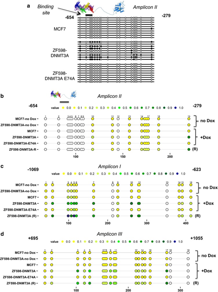
With the recent comprehensive mapping of cancer genomes, there is now a need for functional approaches to edit the aberrant epigenetic state of key cancer drivers to reprogram the epi-pathology of the disease. In this study we utilized a programmable DNA-binding methyltransferase to induce targeted incorporation of DNA methylation (DNAme) in the SOX2 oncogene in breast cancer through a six zinc finger (ZF) protein linked to DNA methyltransferase 3A (ZF-DNMT3A). We demonstrated long-lasting oncogenic repression, which was maintained even after suppression of ZF-DNMT3A expression in tumor cells. The de novo DNAme was faithfully propagated and maintained through cell generations even after the suppression of the expression of the chimeric methyltransferase in the tumor cells. Xenograft studies in NUDE mice demonstrated stable SOX2 repression and long-term breast tumor growth inhibition, which lasted for >100 days post implantation of the tumor cells in mice. This was accompanied with a faithful maintenance of DNAme in the breast cancer implants. In contrast, downregulation of SOX2 by ZF domains engineered with the Krueppel-associated box repressor domain resulted in a transient and reversible suppression of oncogenic gene expression. Our results indicated that targeted de novo DNAme of the SOX2 oncogenic promoter was sufficient to induce long-lasting epigenetic silencing, which was not only maintained during cell division but also significantly delayed the tumorigenic phenotype of cancer cells in vivo, even in the absence of treatment. Here, we outline a genome-based targeting approach to long-lasting tumor growth inhibition with potential applicability to many other oncogenic drivers that are currently refractory to drug design.


Figures

Figure 1
Figure 1
Stable downregulation of the SOX2 expression by ZF598-DNMT3A. (a) Schematic illustration of the SOX2 promoter indicating the domain structure of the ZF598-DNMT3A construct, its binding site and the three amplicons analyzed by sodium bisulfite sequencing or MassARRAYs (amplicon I, blue; II, purple and III, green). (b) Quantification of SOX2 mRNA expression by qRT-PCR in MCF7 cells. Cells were stably transduced with empty vector, ZF598-SKD and ZF598-DNMT3A. The expression of the ZF fusion was controlled by addition or removal of doxycycline (Dox); R=Dox removal. Cells were induced with Dox every 48 h and either harvested at 72 h after first induction (+Dox) or removed from Dox and subcultured for 8 days and processed for western blot. Error bars represent standard deviation (s.d.) (***P< 0.001, **P<0.01, *P<0.05). (c) Detection of SOX2 by western blot. The C-terminal Hemagglutinin (HA) tag was used for immunodetection of the ZF proteins. An anti-histone H3 antibody was used as loading control. (d) Cell viability of MCF7 cells assessed by Cell TiterGlo assays. Empty vector, ZF598-SKD and ZF598-DNMT3A-transduced cells were induced with Dox for 48 h and removed from Dox after 72 h (red arrow). P-values between empty vector and ZF598-DNMT3A and between ZF598-SKD and ZF598-DNMT3A were P<0.001 and P<0.05 respectively, at 144 h.
Figure 2
Figure 2
Expression of the ZF598-DNMT3A induces targeted DNA methylation in the SOX2 promoter. (a) Sodium bisulfite-sequencing analysis of DNA derived from MCF7 cells stably transduced with empty vector, ZF598-DNMT3A and ZF598-DNMT3A-E74A mutant and induced with Dox. The analyzed amplicon II expands from −654 to −279 base pairs (bps) upstream the translation start site and includes the 6ZF-binding site (position −598 relative to the translation start site). Gray circles indicate not analyzed methylation values owing to CpGs with high- or low-mass Dalton peaks falling outside the conservative window of reliable detection for the EpiTYPER software, colored circles indicate variously methylated CpGs. (b) MassARRAY analysis of the same amplicon as in (a). Circles indicate the CpG dinucleotides in the amplicon (Color code: yellow=unmethylated CpG to blue=100% methylated CpG). The percentage of meCpG was determined in MCF7 control-transduced cells and in MCF7 cells stably transduced with ZF598-DNMT3A or ZF-598-DNMT3A-E74A in absence of Dox (no Dox), presence of Dox (+Dox) or after Dox removal (R, eight days). (c) MassARRAY analysis of amplicon I (−1069 to −623 bps upstream of the translation start site) in Dox-free conditions (no Dox) and after Dox induction (+Dox) Dox removal (R). (d) MassARRAY analysis of amplicon III (+695 to +1055 bps) downstream of the translation start site, same conditions as above.
Figure 3
Figure 3
The de novo DNA methylation and oncogenic silencing induced by ZF598-DNMT3A are maintained long term in a xenograft model of breast cancer. (a) Timeline of the subcutaneous tumor injections of MCF7 cells in NUDE mice. (b) Time course plot monitoring tumor volumes of empty vector control and ZF598-DNMT3A animals induced (+Dox) and uninduced (−Dox). Left panel: tumor growth of empty vector control animals. Right panel: tumor volumes of animals implanted with ZF598-DNMT3A. (c) Left panel: tumor volumes of ZF598-DNMT3A and control animals at day 29 post induction. Right panel: tumor volumes of ZF598-DNMT3A implanted animals at day 43, when the −Dox control tumors were collected. P-values between groups are indicated.
Figure 4
Figure 4
MassARRAY analysis of tumor DNA shows targeted induction and maintenance of DNA methylation. DNA from ZF598-DNMT3A +Dox and Dox-removal tumors was extracted at the indicated time points and subjected to sodium bisulfite conversion, followed by MassARRAY to detect DNA methylation frequencies. The amplicon I is located −1069 to −623 bps upstream of the translation start site. Each circle represents a CpG dinucleotide. Color code: yellow=unmethylated CpG, blue=100% methylated CpG. Gray circles indicate not analyzed methylation values due to CpGs with high or low mass Dalton peaks falling outside the conservative window of reliable detection for the EpiTYPER software.
Figure 5
Figure 5
Histological and immunofluorescence analyses of tumor sections reveals phenotypic memory. (a) Hematoxylin and Eosin stains of representative ZF598-DNMT3A −Dox, +Dox and Dox-removal tumor sections. Sections of empty vector tumors were extracted 29 days post induction, sections of ZF598-DNMT3A +Dox were sampled after 54 days of Dox induction, and those of Dox-removal samples were harvested 45 days after Dox removal. Pictures were taken at × 20 and a detail of the image is shown. (b) Immunofluorescence on sections of empty vector and ZF598-DNMT3A tumors collected 29 days post induction. The expression of ZF598-DNMT3A (HA-tag, green), SOX2 (red) and the proliferation marker Ki-67 (green, bottom) in +Dox, −Dox and Dox-removal conditions are indicated. Images are taken at × 40 magnification.

References

    1. 1Kouzarides T. Chromatin modifications and their function. Cell 2007; 128: 693–705. - PubMed
    1. 2Beisel C, Paro R. Silencing chromatin: comparing modes and mechanisms. Nat Rev Genet 2011; 12: 123–135. - PubMed
    1. 3Lange UC, Schneider R. What an epigenome remembers. Bioessays 2010; 32: 659–668. - PubMed
    1. 4Sandoval J, Esteller M. Cancer epigenomics: beyond genomics. Curr Opin Genet Dev 2012; 22: 50–55. - PubMed
    1. 5The Cancer Genome Atlas Network. Comprehensive molecular portraits of human breast tumours. Nature 2012; 490: 61–70. - PMC - PubMed

Publication types

Substances