. 2015 Jul;12(1):180-6.
doi: 10.3892/mmr.2015.3351. Epub 2015 Feb 13.

α-Lipoic acid protects against hypoxia/reoxygenation-induced injury in human umbilical vein endothelial cells through suppression of apoptosis and autophagy

Affiliations

α-Lipoic acid protects against hypoxia/reoxygenation-induced injury in human umbilical vein endothelial cells through suppression of apoptosis and autophagy

Jingjing Zhang et al. Mol Med Rep. 2015 Jul.

Abstract

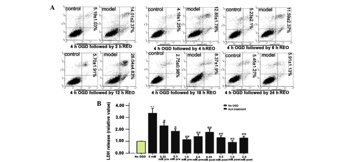
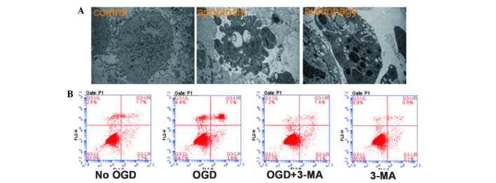
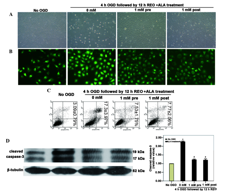
α-Lipoic acid (ALA) is known as a powerful antioxidant, which has been reported to have protective effects against various cardiovascular diseases. The present study aimed to determine whether ALA pre- or post-treatment induced protective effects against hypoxia/reoxygenation-induced injury via inhibition of apoptosis and autophagy in human umbilical vein endothelial cells (HUVECs). In order to simulate the conditions of hypoxia/reoxygenation, HUVECs were subjected to 4 h of oxygen-glucose deprivation (OGD) followed by 12 h of reoxygenation. For the pre-treatment, ALA was added to the buffer 12 h prior to OGD, whereas for the post-treatment, ALA was added at the initiation of reoxygenation. The results demonstrated that ALA pre- or post-treatment significantly reduced lactate dehydrogenase (LDH) release induced through hypoxia/reoxygenation in HUVECs in a dose-dependent manner; of note, 1 mM ALA pre- or post-treatment exhibited the most potent protective effects. In addition, ALA significantly reduced hypoxia/reoxygenation-induced loss of mitochondrial membrane potential, apoptosis and the expression of cleaved caspase-3 in HUVECs. In the presence of the specific autophagy inhibitor 3-methyladenine, hypoxia/reoxygenation-induced apoptosis was significantly reduced. Furthermore, the formation of autophagosomes, cytosolic microtubule-associated protein 1A/1B-light chain 3 ratio and beclin1 levels significantly increased following hypoxia/reoxygenation injury; however, all of these effects were ameliorated following pre- or post-treatment with ALA. The results of the present study suggested that ALA may provide beneficial protection against hypoxia/reoxygenation-induced injury via attenuation of apoptosis and autophagy in HUVECs.


Figures

Figure 1
Figure 1
ALA pre- and post-treatment induces protective effects in HUVECs. (A) Quantification assay for apoptotic activity was evaluated using flow cytometry. HUVECs were exposed to OGD for 4 h followed by reoxygenation for different time periods, with 12 h of reoxygenation inducing the highest levels of apoptosis. Percentages of apoptotic cells (lower right quadrant) as well as apoptotic and necrotic cells (upper right quadrant) are presented as the mean ± standard deviation (n=3). (B) Effect of ALA on LDH release. HUVECs were exposed to OGD for 4 h followed by 12 h of reoxygenation. Pre- or post-treatment with ALA at various concentrations (0.25–2 mM) decreased the OGD-induced increase in LDH release in a concentration-dependent manner. Values are presented as the mean ± standard deviation. **P<0.01 vs. no OGD; #P<0.05 and ##P<0.01 vs. 0 mM ALA treatment. HUVECs, human umbilical vein endothelial cells; OGD, oxygen-glucose deprivation; LDH, lactate dehydrogenase; ALA, α-lipoic acid; REO, reoxygenation.
Figure 2
Figure 2
Hypoxia-induced apoptosis and autophagy in HUVECs. (A) HUVECs were treated with OGD for 4 h followed by 12 h reoxygenation. Apoptosis and autophagy were then observed using electron microscopy (magnification, ×12,000). (B) HUVECs were treated with 3-MA and apoptosis was measured using flow cytometry. Early apoptotic or apoptotic and necrotic cells were identified as single positive for FITC-Annexin V (lower right quadrant) or double positive for both FITC-Annexin V and propidium iodide (upper right quadrant), respectively. HUVECs, human umbilical vein endothelial cells; OGD, oxygen-glucose deprivation; FITC, fluorescein isothiocyanate; 3-MA, 3-methyladenine.
Figure 3
Figure 3
ALA pre- or post-treatment reduces HUVEC apoptosis induced by OGD/reoxygenation. HUVECs were subjected to 4 h of OGD followed by 12 h of reoxygenation in the presence or absence of 1 mM ALA pre- or post-treatment. (A) Cell morphology was observed using inverted phase contrast microscopy (magnification, ×100). (B) Fluorescence microscopy with Rho123 staining was used to detect the mitochondrial membrane potential (magnification, ×400). (C) Cell apoptosis was measured using flow cytometry. Percentages of apoptotic cells (lower right quadrant) as well as apoptotic and necrotic cells (upper right quadrant) are presented as the mean ± standard deviation (n=3). (D) Western blot analysis was used to measure cleaved caspase-3 expression levels and quantitative analysis of these western blots revealed that cleaved caspase-3 was significantly downregulated in ALA pre- or post-treatment groups. *P<0.05 vs. no OGD; #P<0.05 vs. 0 mM ALA treatment. HUVECs, human umbilical vein endothelial cells; OGD, oxygen-glucose deprivation; ALA, α-lipoic acid; REO, reoxygenation.
Figure 4
Figure 4
ALA pre- or post-treatment reduces hypoxia/reoxygenation-induced autophagy in HUVECs. HUVECs were subjected to 4 h of OGD followed by 12 h reoxygenation in the presence or absence of 1 mM ALA pre- or post-treatment. (A) Autophagic vacuoles were stained with monodansylcadaverine, which was detected using fluorescence microscopy (magnification, ×500). (B) Autophagic vacuoles were observed using an electron microscope (magnification upper, 12,000x; lower, 60,000x). Western blot analysis revealed that (C) the conversion of LC3-I to LC3-II was significantly increased in the OGD group and (D) beclin1 was significantly increased in the OGD group, whereas ALA pre- or post-treatment attenuated the effects of hypoxia/reoxygenation-induced injury. *P<0.05 and **P<0.01 vs. no OGD; #P<0.05 vs. 0 mM ALA treatment. HUVECs, human umbilical vein endothelial cells; OGD, oxygen-glucose deprivation; ALA, α-lipoic acid; REO, reoxygenation.

References

    1. Peyrani P, Ramirez J. What is the association of cardiovascular events with clinical failure in patients with community-acquired pneumonia? Infect Dis Clin North Am. 2013;27:205–210. doi: 10.1016/j.idc.2012.11.010. - DOI - PubMed
    1. Navab M, Reddy ST, Van Lenten BJ, Fogelman AM. HDL and cardiovascular disease: atherogenic and atheroprotective mechanisms. Nat Rev Cardiol. 2011;8:222–232. doi: 10.1038/nrcardio.2010.222. - DOI - PubMed
    1. Simbula G, Columbano A, Ledda-Columbano GM, Sanna L, Deidda M, Diana A, Pibiri M. Increased ROS generation and p53 activation in alpha-lipoic acid-induced apoptosis of hepatoma cells. Apoptosis. 2007;12:113–123. doi: 10.1007/s10495-006-0487-9. - DOI - PubMed
    1. Kiffin R, Bandyopadhyay U, Cuervo AM. Oxidative stress and autophagy. Antioxid Redox Signal. 2006;8:152–162. doi: 10.1089/ars.2006.8.152. - DOI - PubMed
    1. Lockshin RA, Zakeri Z. Apoptosis, autophagy and more. Int J Biochem Cell Biol. 2004;36:2405–2419. doi: 10.1016/j.biocel.2004.04.011. - DOI - PubMed

Publication types

MeSH terms