. 2015 Mar 13;458(3):626-631.
doi: 10.1016/j.bbrc.2015.01.158. Epub 2015 Feb 13.

Isoaspartate, carbamoyl phosphate synthase-1, and carbonic anhydrase-III as biomarkers of liver injury

Affiliations

Isoaspartate, carbamoyl phosphate synthase-1, and carbonic anhydrase-III as biomarkers of liver injury

Wayne G Carter et al. Biochem Biophys Res Commun. .

Abstract

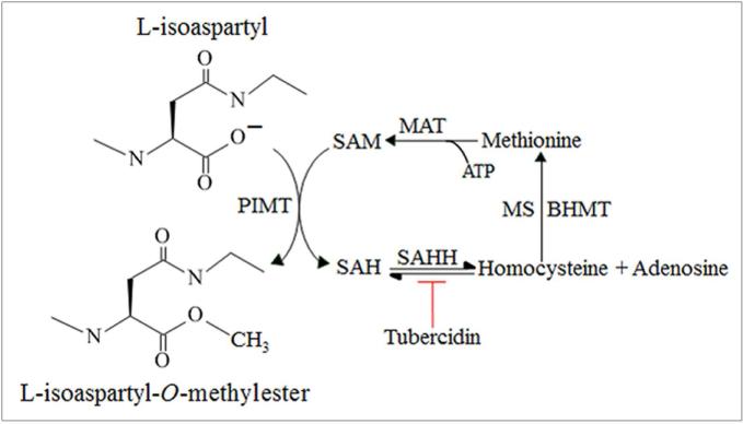
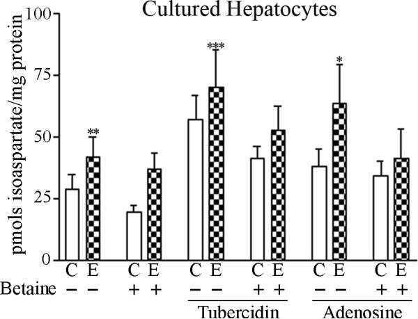
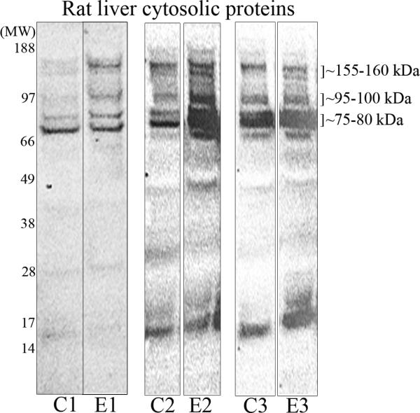
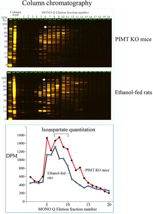
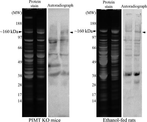
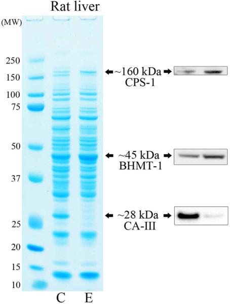
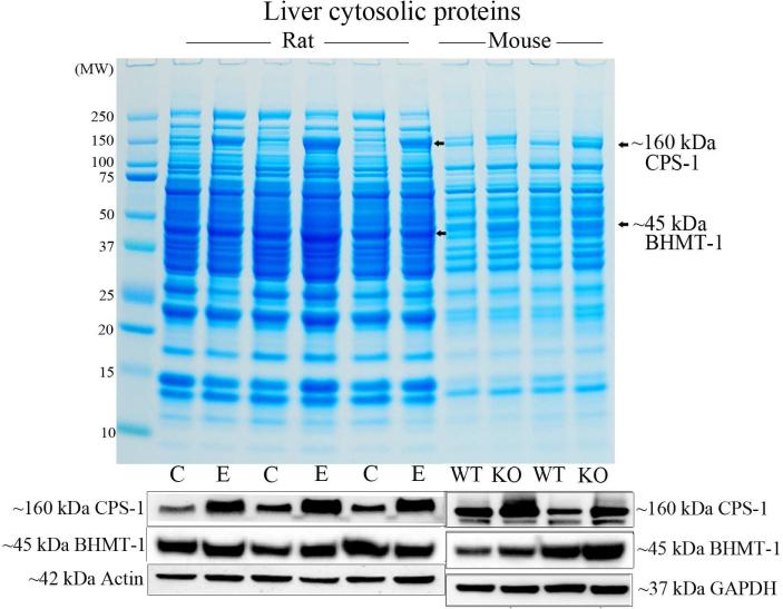
We had previously shown that alcohol consumption can induce cellular isoaspartate protein damage via an impairment of the activity of protein isoaspartyl methyltransferase (PIMT), an enzyme that triggers repair of isoaspartate protein damage. To further investigate the mechanism of isoaspartate accumulation, hepatocytes cultured from control or 4-week ethanol-fed rats were incubated in vitro with tubercidin or adenosine. Both these agents, known to elevate intracellular S-adenosylhomocysteine levels, increased cellular isoaspartate damage over that recorded following ethanol consumption in vivo. Increased isoaspartate damage was attenuated by treatment with betaine. To characterize isoaspartate-damaged proteins that accumulate after ethanol administration, rat liver cytosolic proteins were methylated using exogenous PIMT and (3)H-S-adenosylmethionine and proteins resolved by gel electrophoresis. Three major protein bands of ∼ 75-80 kDa, ∼ 95-100 kDa, and ∼ 155-160 kDa were identified by autoradiography. Column chromatography used to enrich isoaspartate-damaged proteins indicated that damaged proteins from ethanol-fed rats were similar to those that accrued in the livers of PIMT knockout (KO) mice. Carbamoyl phosphate synthase-1 (CPS-1) was partially purified and identified as the ∼ 160 kDa protein target of PIMT in ethanol-fed rats and in PIMT KO mice. Analysis of the liver proteome of 4-week ethanol-fed rats and PIMT KO mice demonstrated elevated cytosolic CPS-1 and betaine homocysteine S-methyltransferase-1 when compared to their respective controls, and a significant reduction of carbonic anhydrase-III (CA-III) evident only in ethanol-fed rats. Ethanol feeding of rats for 8 weeks resulted in a larger (∼ 2.3-fold) increase in CPS-1 levels compared to 4-week ethanol feeding indicating that CPS-1 accumulation correlated with the duration of ethanol consumption. Collectively, our results suggest that elevated isoaspartate and CPS-1, and reduced CA-III levels could serve as biomarkers of hepatocellular injury.

Keywords: Alcohol-induced liver injury; Carbamoyl phosphate synthase-1; Carbonic anhydrase-III; Isoaspartate; Liver proteome; Protein isoaspartyl methyltransferase.


Figures

Figure 1
Figure 1. Carboxyl methylation of isoaspartate protein damage by PIMT
Carboxyl methylation of L-isoaspartyl residues in proteins is catalysed by PIMT using SAM (methyl donor) to form an L-isoaspartyl-O-methylester and SAH. SAH hydrolysis to homocysteine and adenosine is catalysed by SAH hydrolase (SAHH), and can be inhibited by tubercidin. Re-methylation of homocysteine to methionine is provided by either vitamin B12 dependent methionine synthase (MS) activity or from the activity of betaine homocysteine methyltransferase (BHMT) using betaine. The conversion of methionine to SAM by methionine adenosyltransferase (MAT) utilizing ATP completes this cyclic part of the methionine metabolic pathway.
Figure 2
Figure 2. Isoaspartate protein damage in cultured hepatocytes, and rat liver cytosolic proteins
(A) Hepatocytes cultured from control or ethanol-fed rats were treated with exogenous betaine, tubercidin, or adenosine, and the levels of isoaspartate damage quantified. (B) Liver cytosolic proteins from control (C) or ethanol (E) fed rats were methylated by PIMT using 3H-SAM. Radiolabelled proteins were resolved by 1D PAGE and visualised by autoradiography. Increased methylation of proteins from ethanol-fed rats was evident at molecular weights of ~75-80 kDa, ~95-100 kDa, and ~155-160 kDa. Profiles of radiolabelled proteins are shown from 3 different pairs of control and ethanol pair-fed animals.
Figure 3
Figure 3. Enrichment and partial purification of isoaspartate damaged proteins
(A) Liver cytosolic proteins from PIMT KO mice or ethanol-fed rats were enriched through binding and elution from a MONO Q column. Proteins were stained with colloidal Coomassie (upper panel). The start of elution of the ~160 kDa protein doublet in fraction 7 is marked with an arrowhead. The level of isoaspartate damage in each fraction was quantified (lower panel). (B) Peak column fractions 7-10 were concentrated, fractioned, and then resolved by 1D PAGE. Proteins were stained with colloidal Coomassie, or radiolabelled by PIMT using 3H-SAM, and radiolabelled proteins visualised by autoradiography. Co-distribution of protein staining and isoaspartate radiolabelling was evident for the ~160 kDa protein for both PIMT KO mice and ethanol-fed rats (marked with arrowheads).
Figure 4
Figure 4. Comparison of the liver proteome of ethanol-fed rats and PIMT KO mice
(A) Liver proteome from rats fed a control (C) or ethanol (E) diet for 4 weeks were resolved by 1D PAGE and stained with colloidal Coomassie. Elevated CPS-1 and BHMT-1, and reduced CA-III protein levels were visualised by Western blotting. (B) Liver proteome from rats fed a control (C) or ethanol (E) diet for 8 weeks were compared with the proteome of PIMT wild-type (WT) and knockout (KO) mice (upper panel). CPS-1 and BHMT-1 protein levels were visualised by Western blotting (lower panels).

References

    1. Rehm J, Baliunas D, Borges GL, Graham K, Irving H, Kehoe T, Parry CD, Patra J, Popova S, Poznyak V, Roerecke M, Room R, Samokhvalov AV, Taylor B. The relation between different dimensions of alcohol consumption and burden of disease: an overview. Addiction. 2010;105:817–843. - PMC - PubMed
    1. Shield KD, Gmel G, Kehoe-Chan T, Dawson DA, Grant BF, Rehm J. Mortality and potential years of life lost attributable to alcohol consumption by race and sex in the United States in 2005. PLoS One. 2013;8:e51923. - PMC - PubMed
    1. Global status report on alcohol and health 2014. World Health Organization; Geneva: 2014. http://www.who.int/substance_abuse/publications/global_alcohol_report/en/
    1. Cederbaum AI, Lu Y, Wu D. Role of oxidative stress in alcohol-induced liver injury. Arch Toxicol. 2009;83:519–548. - PubMed
    1. Manzo-Avalos S, Saavedra-Molina A. Cellular and mitochondrial effects of alcohol consumption. Int J Environ Res Public Health. 2010;7:4281–4304. - PMC - PubMed

Publication types

MeSH terms