. 2015 Mar 5;57(5):873-886.
doi: 10.1016/j.molcel.2015.01.014. Epub 2015 Feb 12.

Inhibition of Pro-apoptotic BAX by a noncanonical interaction mechanism

Affiliations

Inhibition of Pro-apoptotic BAX by a noncanonical interaction mechanism

Lauren A Barclay et al. Mol Cell. .

Abstract

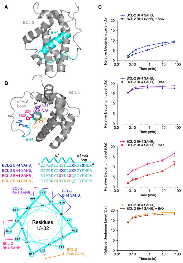
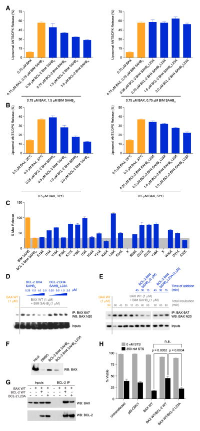
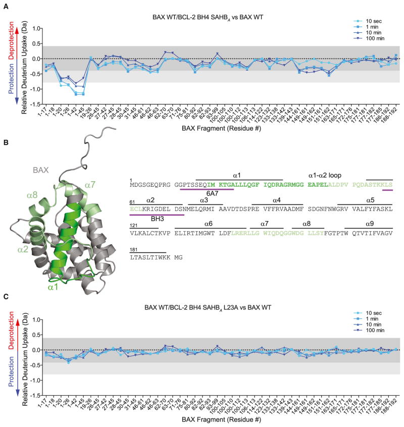
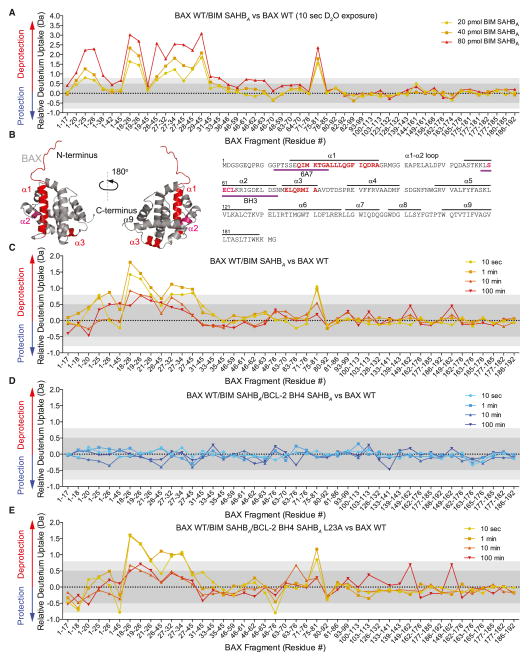
BCL-2 is a negative regulator of apoptosis implicated in homeostatic and pathologic cell survival. The canonical anti-apoptotic mechanism involves entrapment of activated BAX by a groove on BCL-2, preventing BAX homo-oligomerization and mitochondrial membrane poration. The BCL-2 BH4 domain also confers anti-apoptotic functionality, but the mechanism is unknown. We find that a synthetic α-helical BH4 domain binds to BAX with nanomolar affinity and independently inhibits the conformational activation of BAX. Hydrogen-deuterium exchange mass spectrometry demonstrated that the N-terminal conformational changes in BAX induced by a triggering BIM BH3 helix were suppressed by the BCL-2 BH4 helix. Structural analyses localized the BH4 interaction site to a groove formed by residues of α1, α1-α2 loop, and α2-α3 and α5-α6 hairpins on the BAX surface. These data reveal a previously unappreciated binding site for targeted inhibition of BAX and suggest that the BCL-2 BH4 domain may participate in apoptosis blockade by a noncanonical interaction mechanism.


Figures

Figure 1
Figure 1. Sequence, Secondary Structure, and BAX-Binding Activity of BCL-2 BH4 SAHBs
(A) The BH4 domain of BCL-2 comprises α1 and proximal residues of the α1–α2 loop, based on the structure of a BCL-2/BCL-XL chimera (PDB: 1G5M). (B) A panel of stabilized α-helices of BCL-2 domains (SAHBs) was designed based on the BH4 sequence and differential placement of all-hydrocarbon staples (X) along the BH4 domain surfaces. B represents norleucine, which replaced the native methionine to avoid sulfur-based interference with the efficiency of the Grubb’s catalyst. An idealized helical wheel demonstrates the differential placement of all-hydrocarbon staples along the BH4 sequence. (C) The secondary structural stability of BCL-2 BH4 SAHBs in solution as measured by the relative level of deuterium incorporation over time (solid lines) using mass spectrometry. Changes in the rate or overall level of deuterium incorporation by SAHBs in the presence of added recombinant BAX (dashed lines) reflect protein interaction with or without induced peptide folding. Error bars represent the spread of data for all charge states in duplicate experiments. See also Figure S1.
Figure 2
Figure 2. Direct and Sequence-Specific Inhibition of Pro-Apoptotic BAX by the BCL-2 BH4 Domain
(A and B) BCL-2 BH4 SAHBA dose-responsively impaired BIM SAHBA-triggered (A) and heat-induced (B) BAX-mediated liposomal release. L23A mutagenesis of BCL-2 BH4 SAHBA disrupted the BAX-inhibitory activity. Data are mean ± SD for assays performed in triplicate. (C) Alanine scanning was used to identify amino acid residues most critical to functional inhibition of BAX-mediated liposomal release by BCL-2 BH4 SAHBA. Data are mean ± SD for assays performed in triplicate. (D) BCL-2 BH4 SAHBA, but not the L23A mutant, dose-responsively blocked BIM SAHBA-induced exposure of the N-terminal activation epitope on BAX in the presence of liposomes, as assessed by immunoprecipitation with the conformation-specific 6A7 antibody. The data are representative of three independent experiments. (E) Induction of 6A7 epitope exposure by BIM SAHBA in the presence of liposomes was reversed by subsequent treatment (15, 30, and 45 min after BIM SAHBA exposure) with BCL-2 BH4 SAHBA, but not BCL-2 BH4 SAHBA L23A. The data are representative of three independent experiments. (F) L23A mutagenesis of the hydrophobic surface of BCL-2 BH4 SAHBA impaired its interaction with native BAX, as demonstrated by streptavidin pull-down of biotinylated BCL-2 BH4 SAHBs from HeLa cell lysates and anti-BAX western analysis. (G) Co-immunoprecipitation of full-length BCL-2 and BAX co-expressed in HeLa cells was impaired by L23A mutagenesis of the BCL-2 BH4 domain. (H) Correspondingly, L23A mutagenesis reduced the capacity of BCL-2 to protect against staurosporine-induced apoptosis of HeLa cells co-expressing full-length BAX and BCL-2. Data are mean ± SD for experiments performed in triplicate and repeated three times with similar results. See also Figure S2.
Figure 3
Figure 3. Conformational Stabilization of Monomeric BAX by BCL-2 BH4 SAHBA
(A and B) BCL-2 BH4 SAHBA-bound BAX manifests a decrease in deuterium incorporation over time compared to unliganded BAX, as measured by HXMS. The most notable localized protection involves N-terminal fragments that comprise portions of the 6A7 epitope and α1–α2 loop (dark green). Significant, but less prominent, protection was also observed in regions that surround the α1–α2 loop and N terminus (light green). The relative difference plot reflects the relative deuterium incorporation of BCL-2 BH4 SAHBA/BAX WT minus the relative deuterium incorporation of BAX WT. Dark gray shading represents changes in the plot that are below the significance threshold of 0.4 Da, whereas light gray shading and the white region highlight changes above the baseline significance threshold of 0.4 Da and the more stringent threshold of 0.8 Da, respectively. The experiments were repeated twice with similar results. (C) The sequence dependence of BCL-2 BH4 SAHBA-induced conformational stabilization of BAX was confirmed by L23A mutagenesis. BCL-2 BH4 SAHBA L23A had little to no effect on deuterium incorporation by BAX over time. The relative difference plot reflects the relative deuterium incorporation of BCL-2 BH4 SAHBA L23A/BAX WT minus the relative deuterium incorporation of BAX WT. See also Figure S3.
Figure 4
Figure 4. BCL-2 BH4 SAHBA Blocks the Conformational Changes of BAX Induced by BIM SAHBA in a Membrane Environment
(A) The addition of BIM SAHBA to BAX (40 pmol) in a liposomal environment triggered dose-responsive and regiospecific increases in deuterium incorporation compared to unliganded BAX, as measured by HXMS. The most notable region of deprotection involved N-terminal peptides that comprise portions of the 6A7 activation epitope. Significant deprotection was also observed in BAX α3, which comprises a subregion of the C-terminal hydrophobic groove, and in the BH3 domain (BAX α2). The relative difference plot reflects the relative deuterium incorporation of BIM BH3 SAHBA/BAX WT minus the relative deuterium incorporation of BAX WT. Dark gray shading represents changes in the plot that are below the significance threshold of 0.5 Da, whereas light gray shading and the white region highlight changes above the baseline significance threshold of 0.5 Da and the more stringent threshold of 0.8 Da, respectively. Data represent the average of at least two independent experiments. (B) The regions of increased deuterium uptake are highlighted in red (> 0.8 Da significance threshold) and pink (> 0.5 Da significance threshold) on the sequence and structure of monomeric BAX (PDB: 1F16). (C) Incubation of BIM SAHBA (40 pmol) and BAX (40 pmol) in a liposomal environment triggered time-responsive changes in deuterium incorporation that mirrored the regiospecific changes observed upon BIM SAHBA dose escalation. The relative difference plot reflects the relative deuterium incorporation of BIM BH3 SAHBA/BAX WT minus the relative deuterium incorporation of BAX WT. (D) The addition of BCL-2 BH4 SAHBA completely eliminated the observed increases in BAX deuterium exchange triggered by BIM SAHBA in the membrane environment. The relative difference plot reflects the relative deuterium incorporation of BIM BH3 SAHBA/BCL-2 BH4 SAHBA/BAX WT minus the relative deuterium incorporation of BAX WT. (E) The mutant BCL-2 BH4 SAHBA L23A construct had no inhibitory effect, highlighting the peptide sequence specificity of BCL-2 BH4 SAHBA activity. The relative difference plot reflects the relative deuterium incorporation of BIM BH3 SAHBA/BCL-2 BH4 SAHBA L23A/BAX WT minus the relative deuterium incorporation of BAX WT. See also Figure S3.
Figure 5
Figure 5. Localization of the BCL-2 BH4 Binding Region on BAX by Photoaffinity Labeling and Mass Spectrometry
(A and B) Biotinylated BCL-2 BH4 photoreactive SAHBs (pSAHBs) were generated by incorporation of a UV-active benzophenone residue (U) at position 19 (purple) in pSAHBA or position 30 (red) in pSAHBC. pSAHBs were mixed with full-length BAX and subjected to UV irradiation, electrophoresis, excision of the crosslinked protein, trypsin proteolysis, and LC-MS/MS analysis. The plots (above) depict the frequency of crosslinked sites identified across the BAX polypeptide sequence. As a reference, the regions of the BH3-activation sites at the N- and C-terminal faces of BAX are shaded in lavender and teal, respectively, and the C-terminal α9 helix is colored in yellow. The crosslinked residues are mapped onto the solution structure of monomeric BAX (PDB: 1F16) (below) and colored according to the frequency of occurrence. B, norleucine; U, 4-benzoyl-phenylalanine (Bpa). See also Figure S4.
Figure 6
Figure 6. NMR Analysis of the BCL-2 BH4 SAHBA/BAX Interaction
(A–D) NMR analysis of 15N-BAX upon titration with BCL-2 BH4 SAHBA Y28C-MTSLred up to a ratio of 1:1.1 BAX:BCL-2 BH4 SAHB revealed chemical shift changes that localized to the α1–α2 loop, α2, the α2–α3, α3–α4, and α5–α6 hairpins, α8, and α9. Cα atoms of affected residues are represented as orange bars in the plot (A) and orange spheres in the ribbon diagrams (B)–(D). The significance threshold of > 0.043 ppm for backbone amide chemical shift changes was calculated based on the average chemical shift across all residues plus the SD, as previously reported (Gavathiotis et al., 2008) and in accordance with standard methods (Marintchev et al., 2007). As a reference, the α1/α6 region that comprises the N-terminal trigger site is colored lavender, those portions of α-helices 2, 3, 4, and 5 that contain residues forming the C-terminal canonical groove are colored teal, and the C-terminal α9 helix is colored yellow. (E–H) Ratios of BAX cross-peak intensities in the presence of oxidized or reduced (Iox/Ired) BCL-2 BH4 SAHBA Y28C-MTSL plotted versus BAX residue number. BAX residue intensities reduced below a ratio of 0.7 are colored purple in the plot (E) and mapped onto surface views of the BAX structure (F)–(H). BAX residues affected by the MTSL label colocalize to the bottom face of BAX in a region spanning from the base of the C-terminal pocket to the α1–α2 loop of the N-terminal face. See also Figures S5 and S6.
Figure 7
Figure 7. A Distinct Binding Site for BCL-2 BH4 Inhibition of BAX
(A) Docking calculations based on the PRE NMR data placed the BCL-2 BH4 helix at an interaction site formed by the confluence of surface residues from the α1 C terminus, α1–α2 loop, and α2–α3 and α5–α6 hairpins. Those residues within 2 Å of the docked BCL-2 BH4 helix (cyan cylinder) are colored in orange on the ribbon structure of BAX. (B) Surface view of the BH4-interaction site demonstrates a groove comprised of hydrophobic residues (yellow), which are surrounded by a series of positively charged (blue), negatively charged (red), and polar (green) residues. (C) A structural overlay demonstrates that vMIA (aa 131–150) (PDB: 2LR1) and BCL-2 BH4 (aa 13–32) peptides engage BAX at inhibitory sites at the bottom face of BAX.

References

    1. Bird GH, Bernal F, Pitter K, Walensky LD. Synthesis and biophysical characterization of stabilized alpha-helices of BCL-2 domains. Methods Enzymol. 2008;446:369–386. - PMC - PubMed
    1. Braun CR, Mintseris J, Gavathiotis E, Bird GH, Gygi SP, Walensky LD. Photoreactive stapled BH3 peptides to dissect the BCL-2 family interactome. Chem Biol. 2010;17:1325–1333. - PMC - PubMed
    1. Czabotar PE, Westphal D, Dewson G, Ma S, Hockings C, Fairlie WD, Lee EF, Yao S, Robin AY, Smith BJ, et al. Bax crystal structures reveal how BH3 domains activate Bax and nucleate its oligomerization to induce apoptosis. Cell. 2013;152:519–531. - PubMed
    1. Ding J, Mooers BH, Zhang Z, Kale J, Falcone D, McNichol J, Huang B, Zhang XC, Xing C, Andrews DW, Lin J. After embedding in membranes antiapoptotic Bcl-XL protein binds both Bcl-2 homology region 3 and helix 1 of proapoptotic Bax protein to inhibit apoptotic mitochondrial permeabilization. J Biol Chem. 2014;289:11873–11896. - PMC - PubMed
    1. Dlugosz PJ, Billen LP, Annis MG, Zhu W, Zhang Z, Lin J, Leber B, Andrews DW. Bcl-2 changes conformation to inhibit Bax oligomerization. EMBO J. 2006;25:2287–2296. - PMC - PubMed

Publication types

MeSH terms

Substances