. 2015 Feb 16:5:8480.
doi: 10.1038/srep08480.

hiPS-MSCs differentiation towards fibroblasts on a 3D ECM mimicking scaffold

Affiliations

hiPS-MSCs differentiation towards fibroblasts on a 3D ECM mimicking scaffold

Ruodan Xu et al. Sci Rep. .

Abstract

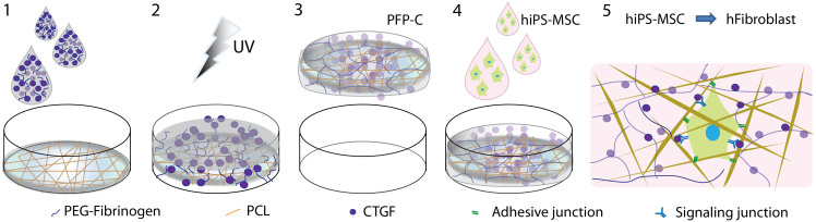
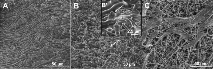
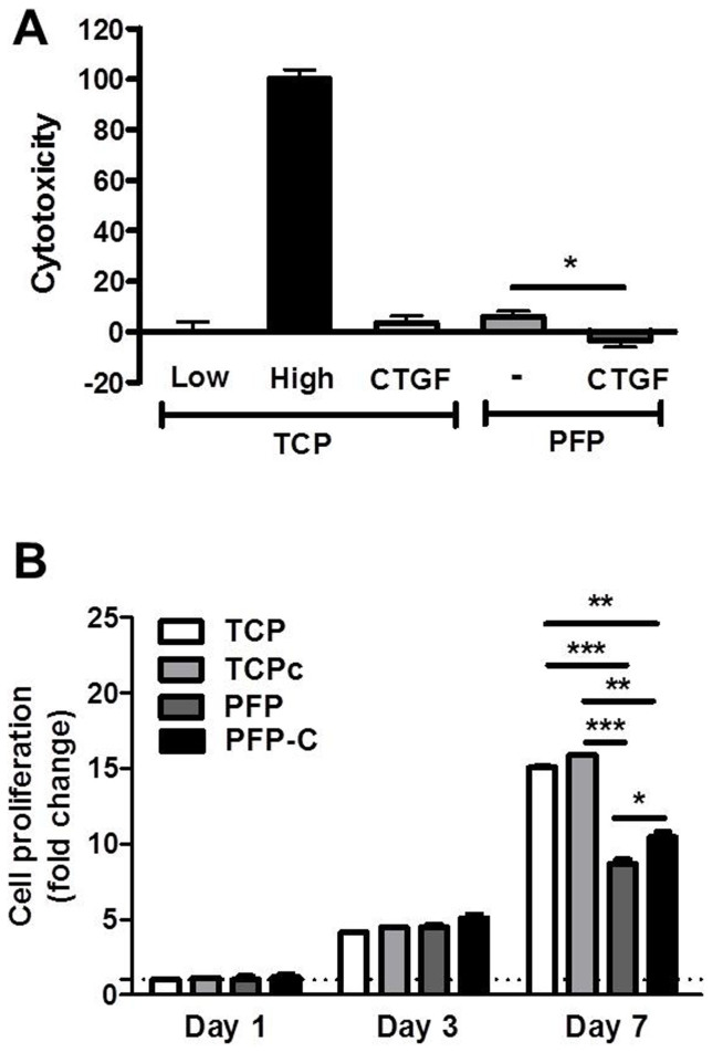
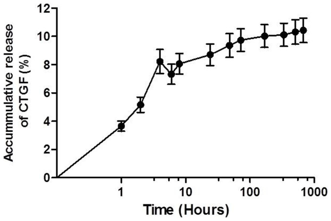
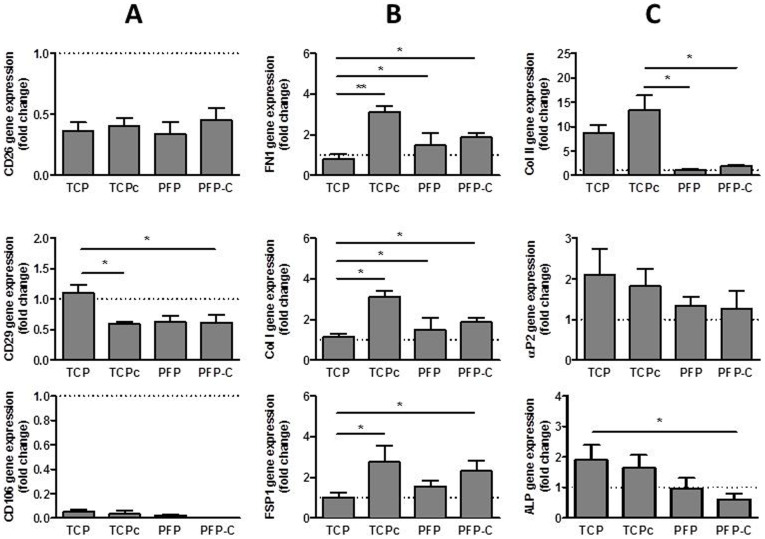
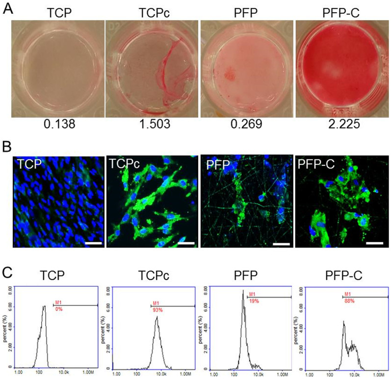
Fibroblasts are ubiquitous cells that constitute the stroma of virtually all tissues and play vital roles in homeostasis. The poor innate healing capacity of fibroblastic tissues is attributed to the scarcity of fibroblasts as collagen-producing cells. In this study, we have developed a functional ECM mimicking scaffold that is capable to supply spatial allocation of stem cells as well as anchorage and storage of growth factors (GFs) to direct stem cells differentiate towards fibroblasts. Electrospun PCL fibers were embedded in a PEG-fibrinogen (PF) hydrogel, which was infiltrated with connective tissue growth factor (CTGF) to form the 3D nanocomposite PFP-C. The human induced pluripotent stem cells derived mesenchymal stem cells (hiPS-MSCs) with an advance in growth over adult MSCs were applied to validate the fibrogenic capacity of the 3D nanocomposite scaffold. The PFP-C scaffold was found not only biocompatible with the hiPS-MSCs, but also presented intriguingly strong fibroblastic commitments, to an extent comparable to the positive control, tissue culture plastic surfaces (TCP) timely refreshed with 100% CTGF. The novel scaffold presented not only biomimetic ECM nanostructures for homing stem cells, but also sufficient cell-approachable bio-signaling cues, which may synergistically facilitate the control of stem cell fates for regenerative therapies.


Figures

Figure 1
Figure 1. Schematic diagram of preparation of PFP-C nanocomposites and their fibrogenic capacity on hiPS-MSCs.
(1) a PF solution with CTGF was added on the top of a PCL mesh; (2) UV light exposure to crosslink PF; (3) a free-standing PFP-C composite was formed; (4) hiPS-MSCs were seeded on the composite; (5) fibrogenesis process synergetically promoted by the adhesive motif on PFP and the signaling induction of CTGF.
Figure 2
Figure 2
Cross-sectional SEM images of PCL fibers alone (A) and PFP (B, B′) composites, arrows indicate the hydrogel component in the composites.(C) SEM image of hiPS-MSCs seeded on the PFP composite.
Figure 3
Figure 3. Biocompatibility of nanocomposites PFP and PFP-C with hiPS-MSCs.
(A) LDH activity measured from the culture media collected 24 hour after hiPS-MSCs seeding on PFP scaffold and PFP-C scaffold. Low toxicity control (0%) was from cells seeded on tissue culture plastic (TCP). High toxicity control (100%) was from cells seeded on TCP and incubated with 1% Triton X-100. (B) Quantitative analysis of 7 day proliferation indexon TCP, TCPc, PFP and PFP-C scaffolds, data were normalized on day 1 (*P < 0.05, **P < 0.005, ***P < 0.0005).
Figure 4
Figure 4. Cumulative release profile of CTGF from PFP-C nanocomposites determined by ELISA assay.
Figure 5
Figure 5
Real time-PCR gene expression analysis of hMSC surface markers CD26, CD29, CD106 (A), fibroblasts markers FSP1, Col I and FN1(B) and adipogenic marker αP2, chondrogenic marker Col II and early osteogenic marker ALP (C) on day 14.(the breaking lines set at 1 are the gene expression levels of undifferentiated hiPS-MSCs) (*P < 0.05, **P < 0.005).
Figure 6
Figure 6. PFP-C nanocomposites enhance hiPS-MSC differentiation towards fibroblasts determined by (A) Collagen content determined by sirius red staining on day 21, (absorbance values at 540 nm are listed) and (B) immunofluorescence analysis of fibroblast marker FSP1(Green) on day 28, nuclei were stained with Hoechst (Blue).
Scale bar = 50 μm. Nuclei of TCP group have diameter 4.1 ± 0.22 μm, nuclei of TCPc group have diameter of 6.3 ± 0.23 μm. (C) Flow cytometry quantification of FSP1 expressed cells on day 28.

References

    1. Ronchetti I., Boraldi F., Annovi G., Cianciulli P. & Quaglino D. Fibroblast involvement in soft connective tissue calcification. Front Genet 4, 22 (2013). - PMC - PubMed
    1. Wu M. Regulation of extracellular matrix remodeling associated with pelvic organ prolapse. J Exp Clin Med 2, 6 (2010).
    1. Van Eijk F. et al. Tissue engineering of ligaments: a comparison of bone marrow stromal cells, anterior cruciate ligament, and skin fibroblasts as cell source. Tissue Eng 10, 893–903 (2004). - PubMed
    1. Chang H. Y. et al. Diversity, topographic differentiation, and positional memory in human fibroblasts. Proc Natl Acad Sci U S A 99, 12877–82 (2002). - PMC - PubMed
    1. Kwan M. D., Slater B. J., Wan D. C. & Longaker M. T. Cell-based therapies for skeletal regenerative medicine. Hum Mol Genet 17, R93–8 (2008). - PubMed

Publication types

Substances

LinkOut - more resources