. 2015 Mar 3;23(3):472-482.
doi: 10.1016/j.str.2015.01.003. Epub 2015 Feb 12.

A bipartite interaction between Hsp70 and CHIP regulates ubiquitination of chaperoned client proteins

Affiliations

A bipartite interaction between Hsp70 and CHIP regulates ubiquitination of chaperoned client proteins

Huaqun Zhang et al. Structure. .

Abstract

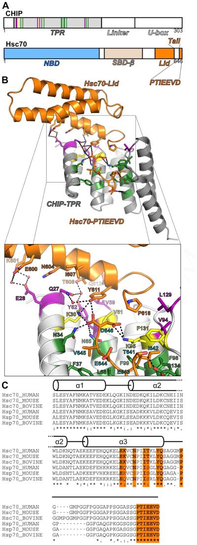
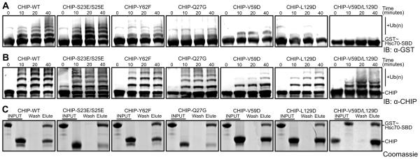
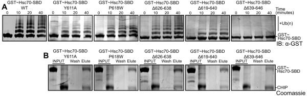
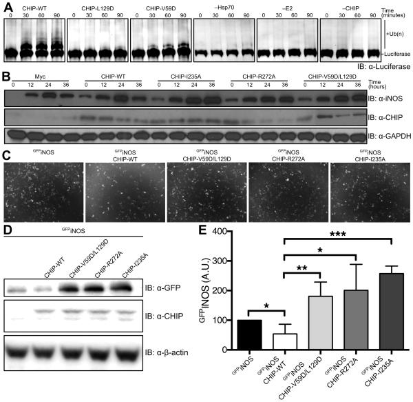
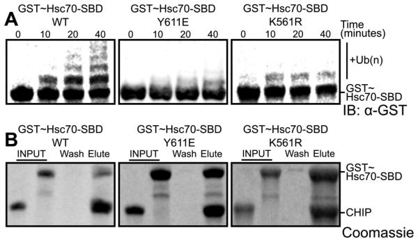
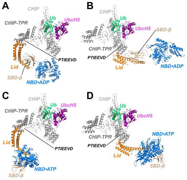
The ubiquitin ligase CHIP plays an important role in cytosolic protein quality control by ubiquitinating proteins chaperoned by Hsp70/Hsc70 and Hsp90, thereby targeting such substrate proteins for degradation. We present a 2.91 Å resolution structure of the tetratricopeptide repeat (TPR) domain of CHIP in complex with the α-helical lid subdomain and unstructured tail of Hsc70. Surprisingly, the CHIP-TPR interacts with determinants within both the Hsc70-lid subdomain and the C-terminal PTIEEVD motif of the tail, exhibiting an atypical mode of interaction between chaperones and TPR domains. We demonstrate that the interaction between CHIP and the Hsc70-lid subdomain is required for proper ubiquitination of Hsp70/Hsc70 or Hsp70/Hsc70-bound substrate proteins. Posttranslational modifications of the Hsc70 lid and tail disrupt key contacts with the CHIP-TPR and may regulate CHIP-mediated ubiquitination. Our study shows how CHIP docks onto Hsp70/Hsc70 and defines a bipartite mode of interaction between TPR domains and their binding partners.


Figures

Figure 1
Figure 1. Structure of the CHIP-TPR/Hsc70-lid-tail Complex
(A) Arrangement of domains within CHIP and Hsc70. (B) Cartoon view of the Hsc70-lid-tail (Hsc70541-646Δ626-638) in complex with the CHIP-TPR domain (CHIP21-154). Hsc70-lid-tail and CHIP-TPR domains are colored orange and grey respectively. Specific CHIP-TPR residues that interact with Hsc70-lid, Hsc70-tail, or both domains are colored green, purple and yellow respectively. (C) Alignment of human, murine and bovine Hsc70- and Hsp70-lid-tail sequences with secondary structure overlay. Lid and tail residues that interact with the CHIP-TPR are colored orange. See also Figures S1, S2 and S3.
Figure 2
Figure 2. Residues that mediate CHIP-TPR/Hsc70-lid interactions modulate CHIP-mediated ubiquitination
(A) Western blot with anti-GST antibody to detect ubiquitination of GST-Hsc70395-646 by wild type (WT) and mutant CHIP1-303. (B) Western blot with anti-CHIP antibody to detect autoubiquitination of wild type (WT) and mutant CHIP1-303. (C) Pull-down of GST~Hsc70395-646 (GST~Hsc70-SBDβ-lid-tail) by wild type (WT) and mutant His6-CHIP1-303 bound to Ni2+ mag-sepharose beads analyzed by SDS PAGE with Coomassie staining. Lanes indicate GST~Hsc70395-646 and CHIP1-303 components utilized (INPUT), the final wash fraction (Wash) and the elution fraction (Elute). See also Figure S4.
Figure 3
Figure 3. Hsc70 residues that mediate CHIP-TPR/Hsc70-lid interactions modulate CHIP-mediated ubiquitination
(A) Western blot with anti-GST antibody to detect ubiquitination of wild type (WT) and mutant GST~Hsc70395-646 (GST~Hsc70-SBDβ-lid-tail) constructs by wild type CHIP1-303. (B) Pull-down of wild type (WT) and mutant GST~Hsc70395-646 constructs by wild type His6-CHIP1-303 bound to Ni2+ mag-sepharose beads analyzed by SDS PAGE with Coomassie staining. Lanes indicate the GST~Hsc70395-646 and CHIP1-303 components utilized (INPUT), the final wash fraction (Wash) and the elution fraction (Elute).
Figure 4
Figure 4. CHIP-mediated Chaperoned Ubiquitination in vitro and ex vivo
(A) CHIP mediated ubiquitination of firefly luciferase was monitored by western blot using an anti-luciferase antibody. Control reactions were carried out with all reagents present except Hsp70, UbcH5b or CHIP respectively. (B) iNOS expression, induced by LPS and IFN-γ treatment, was monitored for 36 hours in Raw264.7 macrophages transfected with wild-type CHIP and CHIP mutants in which interactions between the U-box and E2 enzymes are disrupted (I235A, R272A) or the Hsp70-lid:TPR interaction is disrupted (V59D/L129D). Protein levels were monitored by western blot with anti-iNOS, anti-CHIP and anti-GAPDH (loading control). (C) HEK293 cells were transfected with GFPiNOS and CHIP or CHIP mutants that disrupt E2:U-box interactions (I235A or R272A) or the Hsp70-lid:TPR interaction (V59D/L129D). Cells were incubated for 48 hours prior to imaging. (D) Cells from (C) were then collected, lysed and analyzed by western blot with anti-GFP, anti-CHIP and anti-β-actin (loading control) (E) Western blots from (D) were repeated six times and band intensities were quantified. Data represent mean±SD. *P<0.05. **P<0.01. ***P<0.001.
Figure 5
Figure 5. Posttranslational modifications of Hsc70 residues modulate CHIP-mediated ubiquitination
(A) Western blot with anti-GST antibody to detect ubiquitination of GST~Hsc70395-646Y611E phosphomimic, GST~Hsc70395-646K561R -dimethyl-mimetic in comparison to wild type (WT) GST~Hsc70395-646 by wild type CHIP1-303. (B) Pull-down of GST~Hsc70395-646 Y611E and K561R mutant constructs by wild type His6-CHIP1-303 bound to Ni2+ mag-sepharose beads, analyzed by SDS PAGE with Coomassie staining. Lanes indicate the GST~Hsc70395-646 and CHIP1-303 components utilized (INPUT), the final wash fraction (Wash) and the elution fraction (Elute). See also Figure S5.
Figure 6
Figure 6. Models of Chaperoned Ubiquitination Complexes in ADP- and ATP-Bound States
(A) A model of Hsp70, based on the structure of ADP-bound E. coli DnaK (Bertelsen et al., 2009) is fully compatible with binding to the TPR domain of CHIP protomer with an occluded U-box. CHIP, CHIP-TPR, UbcH5, Ub, Hsc70-Lid, Hsc70-Tail, Hsc70-SBDβ and Hsc70-NBD are colored white, grey, purple, green, orange, black, wheat and blue, respectively. (B) ADP-bound Hsp70 is also compatible with binding to TPR domain of CHIP protomer with accessible U-box. (C) A model of ATP-bound Hsp70, based on the structure of DnaK in the ATP-bound form (Qi et al., 2013), is compatible with binding to CHIP via the TPR domain of the protomer with an occluded U-box. (D) The CHIP-TPR from a protomer with an accessible U-box is also compatible with binding to ATP-bound Hsp70.

References

    1. Adams PD, Afonine PV, Bunkóczi G, Chen VB, Davis IW, Echols N, Headd JJ, Hung L-W, Kapral GJ, Grosse-Kunstleve RW, et al. PHENIX: a comprehensive Python-based system for macromolecular structure solution. Acta Crystallogr. D Biol. Crystallogr. 2010;66:213–221. - PMC - PubMed
    1. Allan RK, Ratajczak T. Versatile TPR domains accommodate different modes of target protein recognition and function. Cell Stress Chaperones. 2011;16:353–367. - PMC - PubMed
    1. Alvira S, Cuéllar J, Röhl A, Yamamoto S, Itoh H, Alfonso C, Rivas G, Buchner J, Valpuesta JM. Structural characterization of the substrate transfer mechanism in Hsp70/Hsp90 folding machinery mediated by Hop. Nat. Commun. 2014;5:5484. - PubMed
    1. Berndsen CE, Wolberger C. New insights into ubiquitin E3 ligase mechanism. Nat. Struct. Mol. Biol. 2014;21:301–307. - PubMed
    1. Bertelsen EB, Zhou H, Lowry DF, Flynn GC, Dahlquist FW. Topology and dynamics of the 10 kDa C-terminal domain of DnaK in solution. Protein Sci. 1999;8:343–354. - PMC - PubMed

Publication types

Associated data

LinkOut - more resources