Review
. 2015 Nov:58:186-210.
doi: 10.1016/j.neubiorev.2015.02.003. Epub 2015 Feb 12.

Toward sophisticated basal ganglia neuromodulation: Review on basal ganglia deep brain stimulation

Affiliations
Review

Toward sophisticated basal ganglia neuromodulation: Review on basal ganglia deep brain stimulation

Claudio Da Cunha et al. Neurosci Biobehav Rev. 2015 Nov.

Abstract

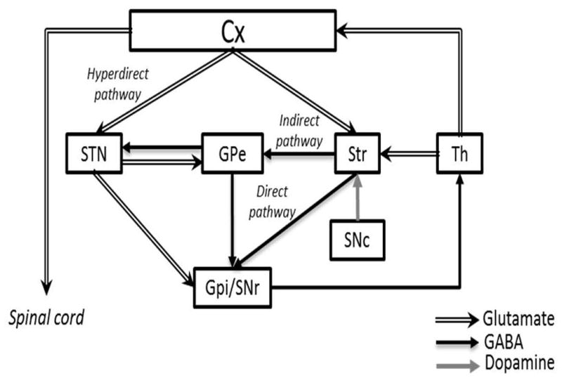
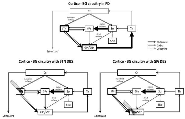
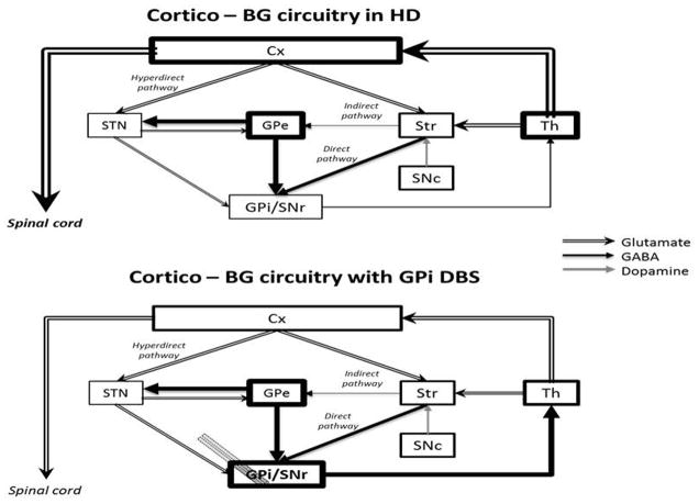
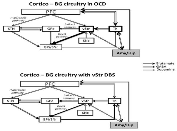
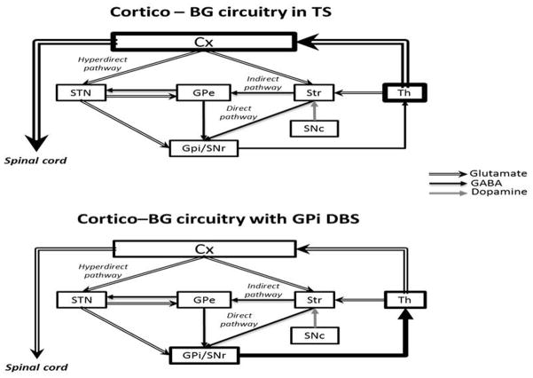
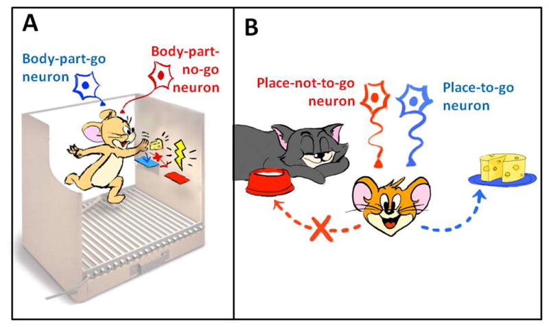
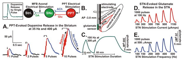
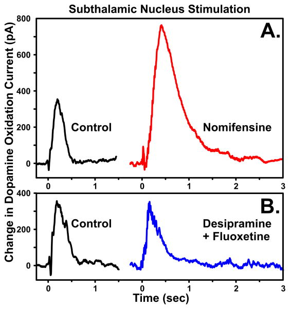
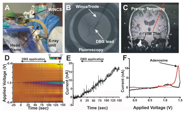
This review presents state-of-the-art knowledge about the roles of the basal ganglia (BG) in action-selection, cognition, and motivation, and how this knowledge has been used to improve deep brain stimulation (DBS) treatment of neurological and psychiatric disorders. Such pathological conditions include Parkinson's disease, Huntington's disease, Tourette syndrome, depression, and obsessive-compulsive disorder. The first section presents evidence supporting current hypotheses of how the cortico-BG circuitry works to select motor and emotional actions, and how defects in this circuitry can cause symptoms of the BG diseases. Emphasis is given to the role of striatal dopamine on motor performance, motivated behaviors and learning of procedural memories. Next, the use of cutting-edge electrochemical techniques in animal and human studies of BG functioning under normal and disease conditions is discussed. Finally, functional neuroimaging studies are reviewed; these works have shown the relationship between cortico-BG structures activated during DBS and improvement of disease symptoms.

Keywords: Deep brain stimulation; Electrochemistry; Functional magnetic resonance imaging; Globus pallidus; Human; Pig; Striatum; Substantia nigra; Subthalamic nucleus; Voltammetry.


Figures

Figure 1
Figure 1
Normal functioning of cortico-BG motor loop as proposed by Alexander, Delong and Strick (1986), Albin, Young, and Penney (1989) and Nambu (2002). Cx, cerebral cortex; GPe, external globus pallidum; GPi, internal globus pallidum; SNc, subtantia nigra pars compacta; SNr, substantia nigra pars reticulata; STN, subthalamic nucleus; Str, striatum; Th, thalamus. Illustration adapted with permission from Nambu (2011).
Figure 2
Figure 2
Hypothetical cortico-BG circuitry functioning in Parkinson’s disease (PD) before and after STN or GPi DBS. Cx, cerebral cortex; GPe, external globus pallidum; GPi, internal globus pallidum; SNc, subtantia nigra pars compacta; SNr, substantia nigra pars reticulata; STN, subthalamic nucleus; Str, striatum; Th, thalamus. Illustration adapted from Nambu (2011).
Figure 3
Figure 3
Hypothetical cortico-BG functioning in Huntington’s disease (HD) before and after GPi DBS. Cx, cerebral cortex; GPe, external globus pallidum; GPi, internal globus pallidum; SNc, subtantia nigra pars compacta; SNr, substantia nigra pars reticulata; STN, subthalamic nucleus; Str, striatum; Th, thalamus. Illustration adapted from Nambu (2011).
Figure 4
Figure 4
Hypothetical cortico-BG circuitry functioning in obsessive compulsive disorder (OCD) before and after ventral striatum DBS. According to this model, obsessions would permanently reverberate in the loop formed by the stimulating activity of the Amy/Hip to Th and then to ventral striatum. The original emotional information would be transmitted through the overactive direct pathway back to Th where it could become a compulsion through thalamocortical activation or return to ventral striatum or Amy/Hip, closing the circuit. Amy, amygdala; GPe, external globus pallidum; GPi, internal globus pallidum; Hip, hippocampus; PFC, prefrontal cortex; SNc, subtantia nigra pars compacta; SNr, substantia nigra pars reticulata; STN, subthalamic nucleus; Str, striatum; vStr, ventral striatum (also as the nucleus accumbens); Th, thalamus. Illustration adapted from Nambu (2011).
Figure 5
Figure 5
Hypothetical cortico-BG circuitry functioning in Tourette syndrome before and after GPi DBS. Cx, cerebral cortex; GPe, external globus pallidum; GPi, internal globus pallidum; SNc, subtantia nigra pars compacta; SNr, substantia nigra pars reticulata; STN, subthalamic nucleus; Str, striatum; Th, thalamus. Illustration adapted from Nambu (2011).
Figure 6
Figure 6
Cartoon illustrating two postulates of the mosaic of broken mirrors model (MBMM). (A) Activation of a striatal “body-part-go cell” selects and initiates a motor action of a body part towards an object. (B) Activation of a striatal “place-to-go cell” selects and initiates the approach to a specific place; activation of a “place-not-to-go cell” prevents approaching to a place.
Figure 7
Figure 7
(A.) DA release in the striatum (Stri.) of urethane anesthetized rats evoked by 1 to 10 pulses of electrical stimulation of pedunculopontine tegmental nucleus (PPT) glutamatergic and cholinergic projections to substantia nigra pars compacta (SNc) DA cells. Note that the increase in DA release is time locked to the stimulation (pulse artifacts are superimposed on the rising portion of the signal) and the recovery to baseline is a reflection of clearance of DA mainly via presynaptic re-uptake. INSET: rapid response of DA to stimulation of DA axons in the medial forebrain bundle (MFB) illustrating that the relatively slower PPT evoked response is trans-synaptically mediated. Glutamate release in the STN evoked by electrical stimulation of the STN at various durations (C.), intensities (D.), and frequencies (E.). (B.) Positioning of a glutamate sensor adjacent to a bipolar stimulating electrode in the STN (see Lee et al., 2007).
Figure 8
Figure 8
Striatal DA is increased with brief STN stimulation. The selectivity of the response was confirmed by (A.) systemic injection of the DA reuptake inhibitor nomifensine (red line), which resulted in a significant increase in DA oxidation current, compared to (B.) serotonin (fluoxetine) and norepinephrine (desipramine) reuptake inhibitors (blue line) which did not significantly increase the STN stimulation-evoked response.
Figure 9
Figure 9
(A.) Represetative example of medial forebrain bundle stimulation-evoked (20 pulses at 100 Hz applied every 10 min) striatal DA release in a urethane anesthetized intact rat and a rat sustaining neurotoxic 6-OHDA lesioning of the nigrostriatal dopaminergic pathway before and following systemic injection of L-DOPA (100 mg/kg i.p.). Note the recovery of stimulated DA release to 100% baseline levels of evoked striatal DA release following administration of L-DOPA in lesioned animals. (B.) Mean ± SEM time courses of effects of L-DOPA injections, compared to saline administration, on striatal DA release in intact and lesioned rats before and following systemic injection of L-DOPA. Solid and dashed blue lines: intact mice that received L-DOPA and saline, respectively. Percentage changes refer to intact pre-saline treated mice.
Figure 10
Figure 10
Intensity, frequency, and site dependency of STN stimulation evoked striatal DA release of the urethane anesthetized rat. (A.) The optimal current intensity for STN stimulation was 300 μA, with the stimulation evoked DA response falling off at 600 μA and greater. (B.) The optimal frequency for STN stimulation was 50 Hz, with the stimulation evoked DA response falling off at 75 Hz and greater. (C.) Prolonged STN stimulation evoked a transient response that peaked within 20 applied pulses (red line), as compared to a 10-fold greater and more sustained DA response to stimulation of the medial forebrain bundle (MFB) (blue line). INSET: DA release evoked by stimulation of the MFB (blue line) was significantly faster at onset compared to STN stimulation when viewed on the same scale as the STN response (red line).
Figure 11
Figure 11
In vivo dopamine (DA) and adenosine (ADO) release measured with WINCS-based FSCV at CFMs in the CN of the isoflurane anesthetized pig. (A.) Electrical stimulation (140 Hz, 0.5 ms- pulse width, for 2 sec) of the STN evoked both DA and adenosine release in the CN. The color plot shows the appearance of DA release immediately during and after stimulation, while the peak corresponding to adenosine release was delayed. (B.) Current versus time plot at +1.5V (blue), +1.0V (red), and +0.6V (black line) shows adenosine first and second oxidation (ADO - 1st, blue and ADO – 2nd, red) and DA (DA, black line) release following electrical stimulation (yellow box). (C.) and (D.) Background subtracted voltammograms for DA and adenosine, respectively, demonstrate simultaneous measurements of DA and adenosine releases (black and blue vertical dashed lines in A).
Figure 12
Figure 12
Intraoperative FSCV recordings during monitoring of tremor in essential tremor patients. (A) FSCV color plot shows increases in oxidation current at +1.4 V (black arrow) and +1.2 V (white arrow) time-locked with the insertion of the DBS electrode into the ventral intermediate nucleus (VIM) of the thalamus (n = 7 patients). (B) Oxidation currents at +1.4 V (black) and +1.2 V (gray) plotted against time. (C) Representative tremor monitoring with a hand- held accelerometer shows tremor was decreased upon DBS electrode insertion. Time scale is the same as in (B). (D) Representative handwriting sample before and after DBS electrode implantation (L, left hand; R, right hand).
Figure 13
Figure 13
Neurochemical changes evoked by DBS in the VIM of the thalamus in patients with essential tremor. (A) Surgical set-up. (B) X-ray of the position of the human CFM and DBS lead in patient’s thalamus. (C) MR image of implantation trajectory. (D) Color plot shows the appearance of oxidation current at +1.4V immediately upon application of DBS (135 Hz, 60 μsec pulse width, 0.5–2.0 V, slowly increased). (E) Current versus time plot at +1.4V following DBS. (F) Background subtracted cyclic voltammogram shows the oxidation peak of adenosine at +1.4 V.

References

    1. Ackermans L, Temel Y, Visser-Vandewalle V. Deep brain stimulation in Tourette’s syndrome. Neurotherapeutics. 2008;5:339–344. - PMC - PubMed
    1. Agnesi F, Tye SJ, Bledsoe JM, Griessenauer CJ, Kimble CJ, Sieck GC, Bennet KE, Garris PA, Blaha CD, Lee KH. Wireless Instantaneous Neurotransmitter Concentration System-based amperometric detection of dopamine, adenosine, and glutamate for intraoperative neurochemical monitoring. J Neurosurg. 2009;111:701–711. - PMC - PubMed
    1. Albin RL, Reiner A, Anderson KD, Penney JB, Young AB. Striatal and nigral neuron subpopulations in rigid Huntington’s disease - implications for the functional-anatomy of chorea and rigidity-akinesia. Ann Neurol. 1990;27:357–365. - PubMed
    1. Albin RL, Young AB, Penney JB. The functional-anatomy of basal ganglia disorders. Trends Neurosci. 1989;12:366–375. - PubMed
    1. Alderson HL, Latimer MP, Blaha CD, Phillips AG, Winn P. An examination of d-amphetamine self-administration in pedunculopontine tegmental nucleus-lesioned rats. Neuroscience. 2004;125:349–358. - PubMed

Publication types