Rheumatoid arthritis affecting temporomandibular joint
- PMID: 25684928
- PMCID: PMC4319332
- DOI: 10.4103/0976-237X.149308
Rheumatoid arthritis affecting temporomandibular joint
Abstract
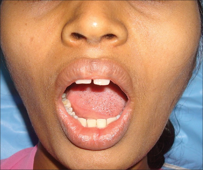
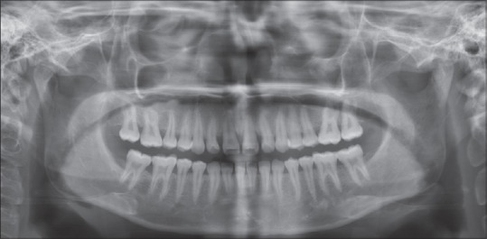
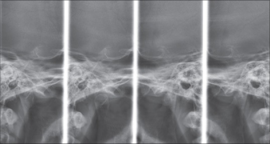
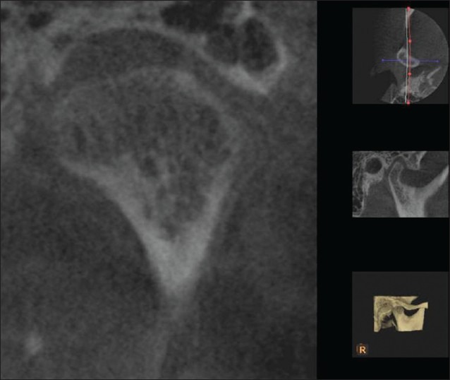
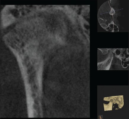
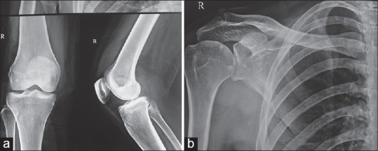
Rheumatoid arthritis (RA) is a chronic, systemic, autoimmune inflammatory disorder that is characterized by joint inflammation, erosive properties and symmetric multiple joint involvement. Temporomandibular joint (TMJ) is very rare to be affected in the early phase of the disease, thus posing diagnostic challenges for the dentist. Conventional radiographs fail to show the early lesions due to its limitations. More recently cone-beam computed tomography (CBCT) has been found to diagnose the early degenerative changes of TMJ and hence aid in the diagnosis of the lesions more accurately. Our case highlights the involvement of TMJ in RA and the role of advanced imaging (CBCT) in diagnosing the bony changes in the early phase of the disease.
Keywords: Cone beam computed tomography; rheumatoid arthritis; temporomandibular joint.
Conflict of interest statement
Figures


References
-
- Chitroda P, Katti G, Ghali S. Bilateral TMJ involvement in rheumatoid arthritis, a case report. J Oral Health Res. 2011;12:74–8.
-
- Milind P, Sushila K. How to live with rheumatoid arthritis. Int Res J Pharm. 2012;3:115–21.
-
- Delantoni A, Spyropoulou E, Chatzigiannis J, Papademitriou P. Sole radiographic expression of rheumatoid arthritis in the temporomandibular joints: A case report. Oral Surg Oral Med Oral Pathol Oral Radiol Endod. 2006;102:e37–40. - PubMed
-
- Seymour RL, Crouse VL, Irby WB. Temporomandibular ankylosis secondary to rheumatoid arthritis. Report of a case. Oral Surg Oral Med Oral Pathol. 1975;40:584–9. - PubMed
-
- Paiva FM, Maria E, Cesar R. Treatment of temporomandibular disorders in children: Summary statements and recommendations. American Academy of Pediatric Dentistry University of Texas Health. Science Center at San Antonio Dental School. J Am Dent Assoc. 1990;120:265–9. - PubMed