. 2015 Feb;29(1):20-9.
doi: 10.1055/s-0035-1544167.

New options for vascularized bone reconstruction in the upper extremity

Affiliations

New options for vascularized bone reconstruction in the upper extremity

Matthew T Houdek et al. Semin Plast Surg. 2015 Feb.

Abstract

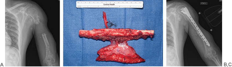
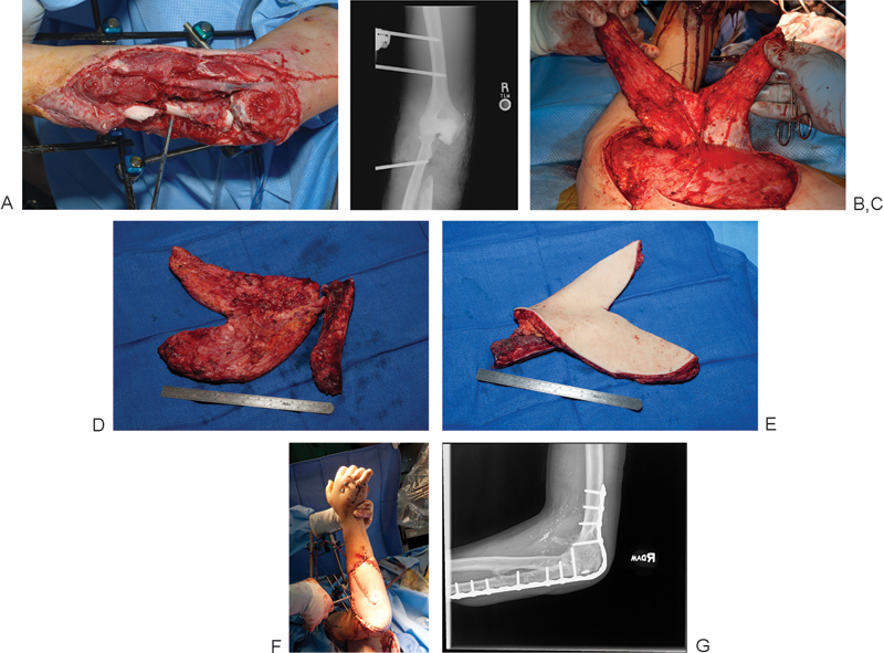
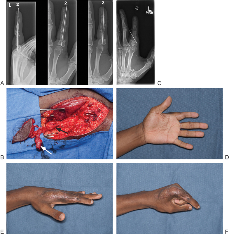
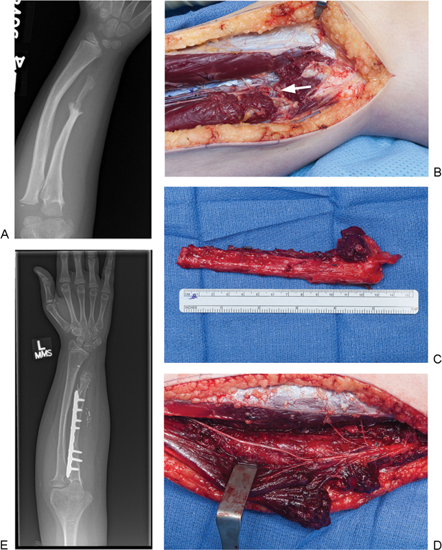
Originally described in the 1970s, vascularized bone grafting has become a critical component in the treatment of bony defects and non-unions. Although well established in the lower extremity, recent years have seen many novel techniques described to treat a variety of challenging upper extremity pathologies. Here the authors review the use of different techniques of vascularized bone grafts for the upper extremity bone pathologies. The vascularized fibula remains the gold standard for the treatment of large bone defects of the humerus and forearm, while also playing a role in carpal reconstruction; however, two other important options for larger defects include the vascularized scapula graft and the Capanna technique. Smaller upper extremity bone defects and non-unions can be treated with the medial femoral condyle (MFC) free flap or a vascularized rib transfer. In carpal non-unions, both pedicled distal radius flaps and free MFC flaps are viable options. Finally, in skeletally immature patients, vascularized fibular head epiphyseal transfer can provide growth potential in addition to skeletal reconstruction.

Keywords: bone graft; medial femoral condylar flap; vascularized bone graft; vascularized epiphyseal transfer; vascularized fibula.


Figures

Fig. 1
Fig. 1
A 26-year-old soldier involved in a land mine accident with segmental humeral loss. (A) Anteroposterior (AP) radiograph of arm showing segmental bone loss in humerus. (B) A vascularized fibula with skin paddle was used as an intercalary graft to reconstruct the humerus. (C) Final AP radiograph showing solid incorporation of the fibular graft.
Fig. 2
Fig. 2
(A) A 19-year-old man who suffered open comminuted fracture of elbow with loss of proximal ulna and concomitant soft tissue loss following an explosion. (B) Anteroposterior (AP) radiograph showing proximal ulnar loss. Both legs were also lost in the injury, making donor sites for vascularized bone limited. (C,D) A composite flap based on the scapular system was designed to incorporate both the parascapular and scapular skin paddles, as well as the bone from the scapular spine. (E) Image of flap prior to inset. (F) Image at time of insetting. (G) Lateral radiograph showing radiographic union.
Fig. 3
Fig. 3
Single- and double-vascularized rib transfers have been found to be successful for clavicle, humerus, and forearm reconstruction. The addition of the serratus muscle provides excellent soft tissue coverage in cases of concomitant soft tissue loss. (A) The technique for elevation involves dissection of the lateral border of the latissimus to expose the serratus. (B) The serratus branch of the thoracodorsal artery is carefully isolated running on the superficial aspect of the serratus (arrow). (C) Two to three slips of the serratus muscle along with two underlying ribs are then dissected. (D) The intercostal muscle is included in the harvest to protect the periosteal blood supply. The chest wall defect is covered with dermal allograft or mesh prior to chest closure.
Fig. 4
Fig. 4
A 35-year-old man suffers a gunshot wound to the index finger resulting in loss of the majority of the proximal phalanx, but maintains finger viability and sensation. (A) Radiographs of finger showing antibiotic impregnated cement spacer placed within proximal phalanx to maintain length. (B) A flap is harvested based on the descending geniculate artery (black arrow) to include bone (white arrow) and skin. Bony healing is obtained in 9 weeks as seen in this (C) anteroposterior (AP) radiograph (D–F) and final function is acceptable.
Fig. 5
Fig. 5
(A) A 9-year-old girl who presents with distal ulnar hypoplasia and non-union. Proximal fibular physeal transfer was used for reconstruction of the involved ulna. (B) Image of perforating branch to physis originating from the anterior tibial artery (arrowhead). (C) Image of graft prior to insetting. (D) Image of donor site showing carefully preserved branches of deep peroneal nerve, which must be preserved during dissection to avoid a postoperative foot drop. (E) Anteroposterior (AP) radiograph of forearm showing proximal bony union and re-establishment of forearm length.

References

    1. Wood M B, Cooney W P III, Irons G B Jr. Skeletal reconstruction by vascularized bone transfer: indications and results. Mayo Clin Proc. 1985;60(11):729–734. - PubMed
    1. Wood M B. Free vascularized bone transfers for nonunions, segmental gaps, and following tumor resection. Orthopedics. 1986;9(6):810–816. - PubMed
    1. de Boer H H, Wood M B. Bone changes in the vascularised fibular graft. J Bone Joint Surg Br. 1989;71(3):374–378. - PubMed
    1. Dell P C, Sheppard J E. Vascularized bone grafts in the treatment of infected forearm nonunions. J Hand Surg Am. 1984;9(5):653–658. - PubMed
    1. Taylor G I, Miller G D, Ham F J. The free vascularized bone graft. A clinical extension of microvascular techniques. Plast Reconstr Surg. 1975;55(5):533–544. - PubMed