. 2015:2015:747245.
doi: 10.1155/2015/747245. Epub 2015 Jan 20.

Mesenchymal Stem Cells Pretreated with HGF and FGF4 Can Reduce Liver Fibrosis in Mice

Affiliations

Mesenchymal Stem Cells Pretreated with HGF and FGF4 Can Reduce Liver Fibrosis in Mice

Sulaiman Shams et al. Stem Cells Int. 2015.

Abstract

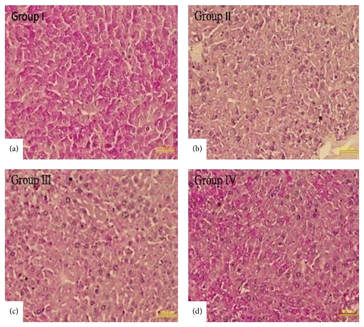
Stem cells have opened a new avenue to treat liver fibrosis. We investigated in vitro and in vivo the effect of cytokine (HGF and FGF4) pretreated MSCs in reduction of CCl4 liver injury. Mouse MSCs were pretreated with cytokines to improve their ability to reduce CCl4 injury. In vitro we gave CCl4 injury to mouse hepatocytes and cocultured it with untreated and cytokines pretreated MSCs. For in vivo study we labeled MSCs with PKH-26 and transplanted them into CCl4 injured mice by direct injection into liver. In vitro data showed that cytokines pretreated MSCs significantly reduce LDH level and apoptotic markers in CCl4 injured hepatocytes cocultured model. Furthermore the cytokines pretreated MSCs also improved cell viability and enhanced hepatic and antiapoptotic markers in injured hepatocytes cocultured model as compared to untreated MSCs. In vivo data in cytokines pretreated group demonstrated greater homing of MSCs in liver, restored glycogen storage, and significant reduction in collagen, alkaline phosphatase, and bilirubin levels. TUNEL assay and real time PCR also supported our hypothesis. Therefore, cytokines pretreated MSCs were shown to have a better therapeutic potential on reduction of liver injury. These results demonstrated the potential utility of this novel idea of cytokines pretreated MSCs for the treatment of liver fibrosis.


Figures

Figure 1
Figure 1
Morphological changes in cytokines pretreated MSCs: (a) MSCs as negative control; (b, c, d, and e) 7-, 15-, 21-, and 28-day cytokines pretreated MSCs; (f) mature hepatocytes as positive control (200x, scale bar ~200 μm).
Figure 2
Figure 2
(a) Expression of hepatic markers: (A1) RT-PCR analysis of MSCs pretreated with cytokines (Lanes 1–5) 0, 7, 15, 21, and 28 days for the expression of Albumin, Cyt-8, and Cyt-18. (Lane 6), mature hepatocytes were used as positive control. (A2) Gel band quantification by Image J software. (b) Immunocytochemistry of cytokines pretreated MSCs. (A–D) Expression of hepatic markers at protein level for Albumin, Cytokeratin 8, Cytokeratin 18, and AFP on day 15, respectively. Nuclei were counterstained with DAPI (blue) (200x, scale bar ~100 μm). (c) PAS staining after pretreatment for glycogen storage: (A, B, C) PAS staining of cytokines pretreated MSCs on days 0, 15, and 21. (D) Mature hepatocytes as positive control (200x, scale bar ~100 μm).
Figure 3
Figure 3
LDH assay of normal hepatocytes, CCl4 injured hepatocytes (3 mM, 5 mM) for 2 hrs, 4 hrs, and 6 hrs.
Figure 4
Figure 4
Effects of cytokines pretreated MSCs in injured hepatocytes cocultured model. (a) LDH release was estimated in the cell supernatant. (b) Cell viability was determined by counting damaged cells using trypan blue exclusion test. LDH release was reduced and the number of damaged cells was decreased in cytokines pretreated group as compared with control and untreated MSCs group. All values were expressed as mean + SEM. * P < 0.05 versus control and untreated MSCs group.
Figure 5
Figure 5
(a) Gene expression profile of MSCs/CCl4 injured hepatocytes coculture model: (Lane 1) normal hepatocytes, (Lane 2) CCl4 injured hepatocytes as control, and (Lanes 3 and 4) CCl4 injured hepatocytes cocultured with untreated and cytokines pretreated MSCs, respectively. (b) Quantification of gel band by image J software: * P < 0.05 versus control and untreated MSCs group.
Figure 6
Figure 6
(a and b) Localization of untreated and cytokines pretreated MSCs in Group III and Group IV animals. (c) Quantification of engrafted cells in Groups III and IV. (100x, scale bar ~100 μm). * P < 0.05 was considered to be significant.
Figure 7
Figure 7
Sirius Red staining in liver section: (a–d) representing Groups I–IV; Sirius Red staining showed reduced collagen level in Group IV as compared to other groups (200x, scale bar ~100 μm). (e) Bar graph represents percentage of fibrosis between the groups: * P was < 0.05 for Group I versus Groups II, III, and IV. # P < 0.05 for Group III versus Group IV.
Figure 8
Figure 8
Functional analysis after MSCs transplantation: (a) bilirubin, (b) alkaline phosphatase; levels in Groups I, II, III, and IV after 3 weeks of transplantation. * P value < 0.05 was considered significant.
Figure 9
Figure 9
PAS staining for the storage of glycogen in liver sections of Group I to Group IV (a–d) (200x, scale bar ~100 μm).
Figure 10
Figure 10
TUNEL apoptotic assay: assessment of apoptosis in experimental groups. (a–d) representing Groups I to IV: TUNEL-positive nuclei (green) in liver sections represent apoptotic cells. Nuclei were counterstained with DAPI (blue) (400x, scale bar ~100 μm).
Figure 11
Figure 11
(a) Gene expression profiling after 3 weeks of transplantation: real time PCR analysis for apoptotic (Bax, caspase-3, NF-κβ, andTNF-α) genes between the experimental groups. The * P < 0.05 versus Group III and Group II. (b) Gene expression profiling after 3 weeks of transplantation: real time PCR analysis for hepatic (Albumin, Cytokeratin 8) and antiapoptotic (Bcl-xl, Bcl 2) genes between the experimental groups. The * P < 0.05 versus Group III and Group II.

References

    1. Friedman S. L. Mechanisms of disease: mechanisms of hepatic fibrosis and therapeutic implications. Nature Clinical Practice: Gastroenterology & Hepatology. 2004;1(2):98–105. - PubMed
    1. Zhao D. C., Lei J. X., Chen R., et al. Bone marrow-derived mesenchymal stem cells protect against experimental liver fibrosis in rats. World Journal of Gastroenterology. 2005;11(22):3431–3440. - PMC - PubMed
    1. Carvalho A. B., Quintanilha L. F., Dias J. V., et al. Bone marrow multipotent mesenchymal stromal cells do not reduce fibrosis or improve function in a rat model of severe chronic liver injury. Stem Cells. 2008;26(5):1307–1314. doi: 10.1634/stemcells.2007-0941. - DOI - PubMed
    1. Liang L., Ma T., Chen W., et al. Therapeutic potential and related signal pathway of adipose-derived stem cell transplantation for rat liver injury. Hepatology Research. 2009;39(8):822–832. doi: 10.1111/j.1872-034x.2009.00506.x. - DOI - PubMed
    1. Rabani V., Shahsavani M., Gharavi M., Piryaei A., Azhdari Z., Baharvand H. Mesenchymal stem cell infusion therapy in a carbon tetrachloride-induced liver fibrosis model affects matrix metalloproteinase expression. Cell Biology International. 2010;34(6):601–605. doi: 10.1042/CBI20090386. - DOI - PubMed

LinkOut - more resources