

In vitro study of nano-hydroxyapatite/chitosan-gelatin composites for bio-applications
- PMID: 25685488
- PMCID: PMC4294712
- DOI: 10.1016/j.jare.2013.02.004
In vitro study of nano-hydroxyapatite/chitosan-gelatin composites for bio-applications
Abstract
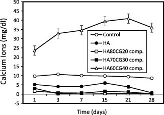
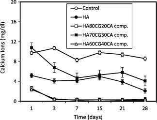
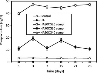
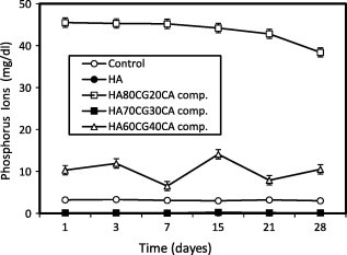
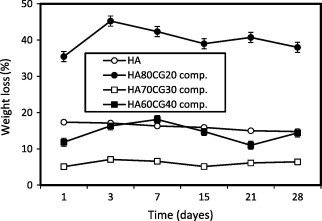
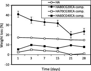
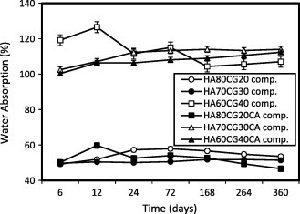
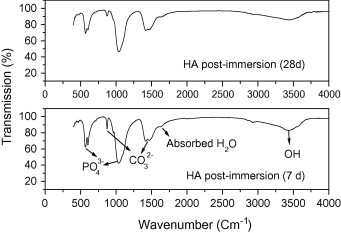
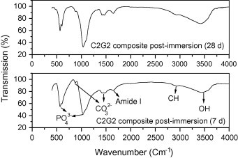
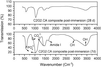
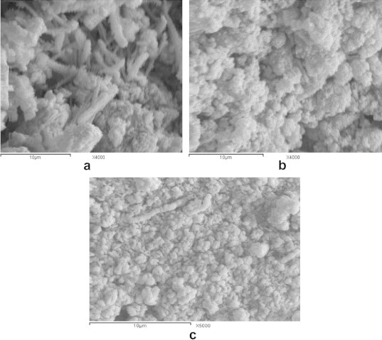
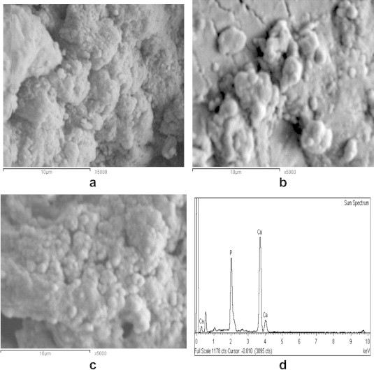
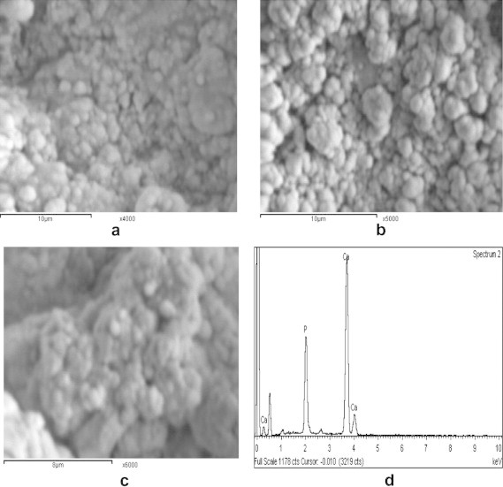
The present work aims to study the in vitro properties of nano-hydroxyapatite/chitosan-gelatin composite materials. In vitro behavior was performed in simulated body fluid (SBF) to verify the formation of apatite layer onto the composite surfaces. The in vitro data proved the deposition of calcium and phosphorus ions onto hydroxyapatite /polymeric composite surfaces especially those containing high concentrations of polymer content. The degradation of the composites decreased with increase in the polymeric matrix content and highly decreased in the presence of citric acid (CA), especially these composites which contain 30% polymeric content. The water absorption of the composites increased with increase in the polymeric content and highly increased with CA addition. The Fourier transformed infrared reflectance (FT-IR) and scanning electron microscope (SEM) for the composites confirmed the formation of bone-like apatite layer on the composite surfaces, especially those containing high content of polymers (30%) with 0.2 M of CA. These promising composites have suitable properties for bio-applications such as bone grafting and bone tissue engineering applications in the future.
Keywords: Chitosan; Composites; Hydroxyapatite; In vitro; SEM.
Figures

Similar articles
-
Physicochemical and biological properties of hydrogel/gelatin/hydroxyapatite PAA/G/HAp/AgNPs composites modified with silver nanoparticles.J Nanosci Nanotechnol. 2012 Dec;12(12):9302-11. doi: 10.1166/jnn.2012.6756. J Nanosci Nanotechnol. 2012. PMID: 23447993
-
Fabrication and mechanical evaluation of hydroxyapatite/oxide nano-composite materials.Mater Sci Eng C Mater Biol Appl. 2013 Oct;33(7):4126-32. doi: 10.1016/j.msec.2013.05.059. Epub 2013 Jun 6. Mater Sci Eng C Mater Biol Appl. 2013. PMID: 23910323
-
The comparison study of bioactivity between composites containing synthetic non-substituted and carbonate-substituted hydroxyapatite.Mater Sci Eng C Mater Biol Appl. 2016 May;62:260-7. doi: 10.1016/j.msec.2016.01.056. Epub 2016 Jan 26. Mater Sci Eng C Mater Biol Appl. 2016. PMID: 26952422
-
Applications of Polymeric Composites in Bone Tissue Engineering and Jawbone Regeneration.Polymers (Basel). 2021 Oct 6;13(19):3429. doi: 10.3390/polym13193429. Polymers (Basel). 2021. PMID: 34641243 Free PMC article. Review.
-
Recent Advances in Applications of Cellulose Derivatives-Based Composite Membranes with Hydroxyapatite.Materials (Basel). 2020 May 29;13(11):2481. doi: 10.3390/ma13112481. Materials (Basel). 2020. PMID: 32486050 Free PMC article. Review.
Cited by
-
Improving the Mechanical Resistance of Hydroxyapatite/Chitosan Composite Materials Made of Nanofibers with Crystalline Preferential Orientation.Materials (Basel). 2022 Jul 5;15(13):4718. doi: 10.3390/ma15134718. Materials (Basel). 2022. PMID: 35806844 Free PMC article.
-
Highly Segregated Biocomposite Membrane as a Functionally Graded Template for Periodontal Tissue Regeneration.Membranes (Basel). 2021 Aug 30;11(9):667. doi: 10.3390/membranes11090667. Membranes (Basel). 2021. PMID: 34564484 Free PMC article.
-
Deeply Implanted Conformal Antenna for Real-Time Bio-Telemetry Applications.Sensors (Basel). 2024 Feb 10;24(4):1170. doi: 10.3390/s24041170. Sensors (Basel). 2024. PMID: 38400327 Free PMC article.
-
Gelatin-polysaccharide composite scaffolds for 3D cell culture and tissue engineering: Towards natural therapeutics.Bioeng Transl Med. 2018 Dec 28;4(1):96-115. doi: 10.1002/btm2.10124. eCollection 2019 Jan. Bioeng Transl Med. 2018. PMID: 30680322 Free PMC article. Review.
-
Chitosan and Its Potential Use as a Scaffold for Tissue Engineering in Regenerative Medicine.Biomed Res Int. 2015;2015:821279. doi: 10.1155/2015/821279. Epub 2015 Oct 4. Biomed Res Int. 2015. PMID: 26504833 Free PMC article. Review.
References
-
- Murugan R., Ramakrishna S. Nano-structured biomaterials. In: Nalwa H.S., editor. Encyclopedia of nanoscience and nanotechnology, vol. 7. American Scientific Publishers; California: 2004. p. 595.
-
- Li J., Pin C.Y., Yuji Y., Yao F., Yao K. Modulation of nano-hydroxyapatite size via formation on chitosan–gelatin network film in situ. Biomaterials. 2007;28:781–790. - PubMed
-
- Kong L., Gao Y., Lu G., Gong Y., Zhao N., Zhang X. A study on the bioactivity of chitosan/nanohydroxyapatite composite scaffolds for bone tissue engineering. Eur Polym J. 2006;42:3171–3179.
-
- Xianmiao C., Yubao L., Yi Z., Li Z., Jidong L., Huanan W. Properties and in vitro biological evaluation of nano-hydroxyapatite/chitosan membranes for bone guided regeneration. Mater Sci Eng C. 2009;29:29–35.
LinkOut - more resources
Full Text Sources
Other Literature Sources
