

doi: 10.1155/2015/467243.
Epub 2015 Jan 19.
A rare combination of ovarian and uterine leiomyomas with goblet cell carcinoid of the appendix
Affiliations
- PMID: 25685587
- PMCID: PMC4313517
- DOI: 10.1155/2015/467243
Item in Clipboard
A rare combination of ovarian and uterine leiomyomas with goblet cell carcinoid of the appendix
Case Rep Surg.
2015.
Abstract
We present a case of the rare combination of unilateral ovarian leiomyoma, uterine leiomyoma, and goblet cell carcinoid tumor of the appendix in a premenopausal woman who presented with right iliac pain. Immunohistochemistry study for desmin (muscle marker) and chromogranin and synaptophysin (neuroendocrine markers) confirmed immunophenotyping origin. Interestingly, both tumors showed positive reaction for estrogen receptor. To our knowledge, such a combination has not been reported previously in the literature. In this paper, the pathogenesis and differential diagnosis of both types of tumors are discussed.
Figures
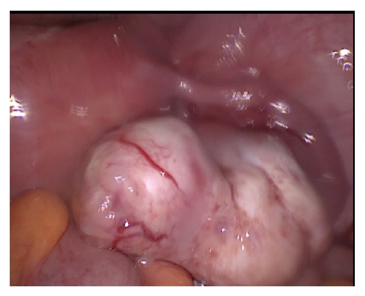
Laparoscopic intraoperative view of the right ovary, right tube, and part of the uterus. The right ovary is noted to be enlarged with a 5 × 3 cm hard mass separated from the uterus not adherent to or infiltrating the surroundings.
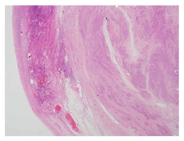
Low power microscopic view showing a normal ovarian tissue in the left side with a well-defined leiomyoma in the right side of the picture (H&E).
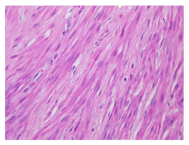
High power view of the ovarian leiomyoma composed of interlacing spindle cells with characteristic cigar-shaped nuclei (H&E).
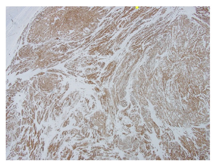
Low microscopic view shows diffuse strong positivity of the ovarian leiomyoma cells for desmin marker confirming smooth cell origin (immunohistochemistry stain).
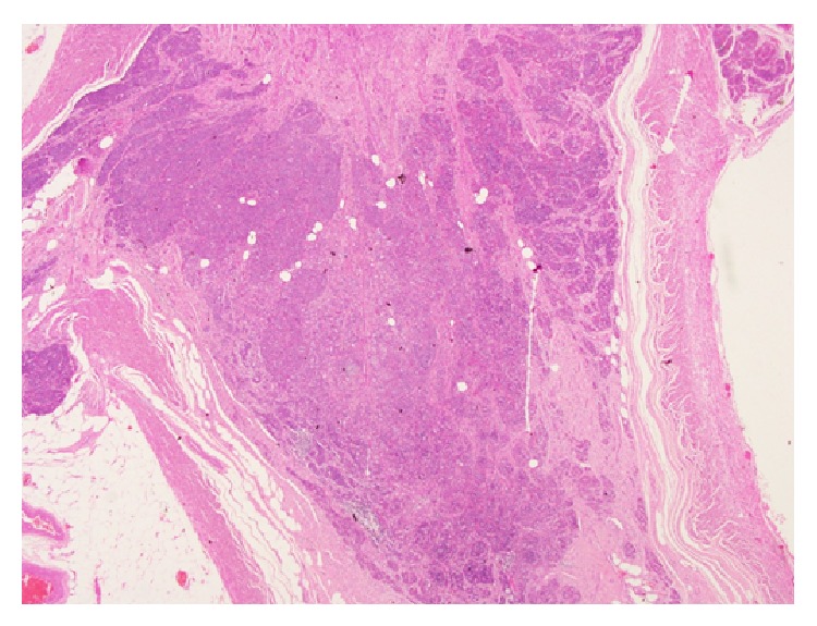
Low power microscopic view of the goblet cell carcinoid involving the entire wall to serosal level of the distal part of the appendix (H&E stained).
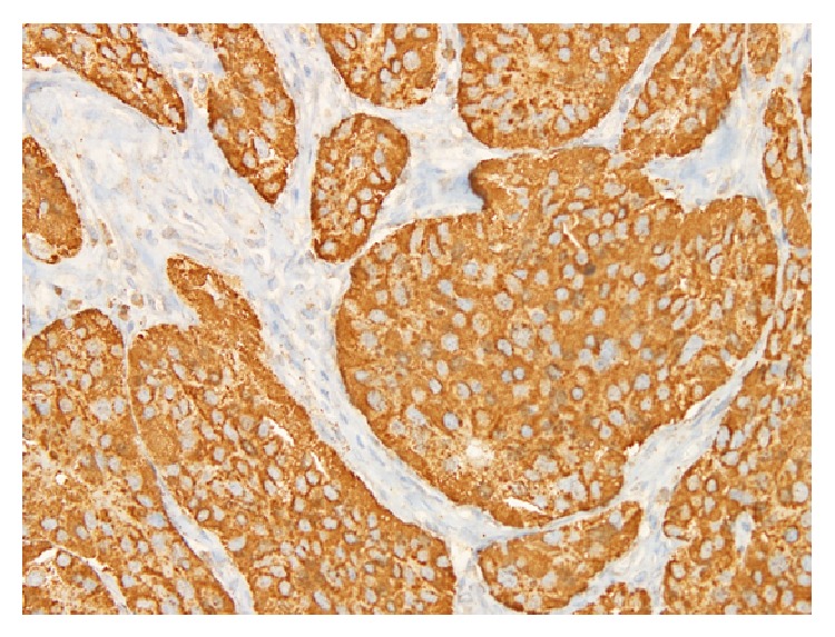
Microscopic view of the goblet cell carcinoid showing strong positive reaction of the tumor for the chromogranin marker (immunohistochemistry stain).
Similar articles
-
Combined classical carcinoid and goblet cell carcinoid tumor: a new morphologic variant of carcinoid tumor of the appendix.Am J Surg Pathol. 2010 Aug;34(8):1163-7. doi: 10.1097/PAS.0b013e3181e52916. Am J Surg Pathol. 2010. PMID: 20631606
-
Lipid-rich and clear cell neuroendocrine tumors ("carcinoids") of the appendix: potential confusion with goblet cell carcinoid.Am J Surg Pathol. 2010 Mar;34(3):401-4. doi: 10.1097/PAS.0b013e3181ce9204. Am J Surg Pathol. 2010. PMID: 20139759
-
Comparative analysis of alternative and traditional immunohistochemical markers for the distinction of ovarian sertoli cell tumor from endometrioid tumors and carcinoid tumor: A study of 160 cases.Am J Surg Pathol. 2007 Feb;31(2):255-66. doi: 10.1097/01.pas.0000213355.72638.f4. Am J Surg Pathol. 2007. PMID: 17255771
-
A subserosal, pedunculated, multilocular uterine leiomyoma with ovarian tumor-like morphology and histological architecture of adenomatoid tumors: a case report and review of the literature.J Med Case Rep. 2016 Dec 20;10(1):352. doi: 10.1186/s13256-016-1167-1. J Med Case Rep. 2016. PMID: 27998309 Free PMC article. Review.
-
Atypical uterine leiomyoma: a case report and review of the literature.J Med Case Rep. 2016 Jan 22;10:22. doi: 10.1186/s13256-016-0800-3. J Med Case Rep. 2016. PMID: 26801982 Free PMC article. Review.
Cited by
-
Case Report: Primary Ovarian Leiomyoma: A Clinical Analysis of Case Series and Literature Review.Front Med (Lausanne). 2022 Apr 1;9:822339. doi: 10.3389/fmed.2022.822339. eCollection 2022. Front Med (Lausanne). 2022. PMID: 35433727 Free PMC article.
References
LinkOut - more resources
Full Text Sources
Other Literature Sources
