. 2014 Nov 3;3(6):e653-60.
doi: 10.1016/j.eats.2014.08.002. eCollection 2014 Dec.

Arthroscopic bone graft procedure for anterior inferior glenohumeral instability

Affiliations

Arthroscopic bone graft procedure for anterior inferior glenohumeral instability

Ettore Taverna et al. Arthrosc Tech. .

Abstract

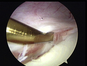
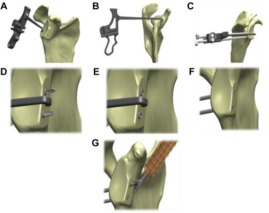
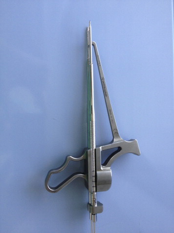
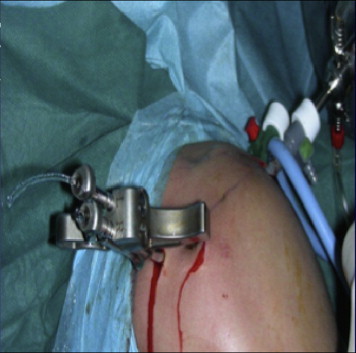
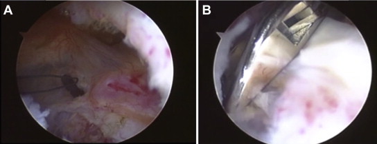
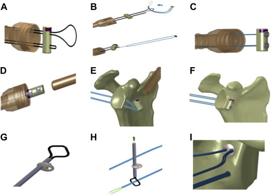
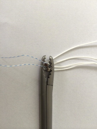
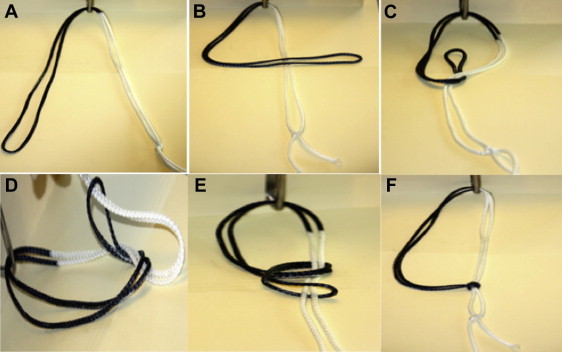
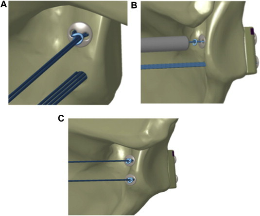
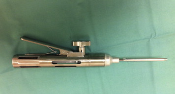
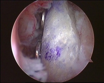
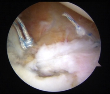
There are many described surgical techniques for the treatment of recurrent anterior shoulder instability. Numerous authors have performed anterior bone block procedures with good results for the treatment of anterior shoulder instability with glenoid bone loss. The benefits of using arthroscopic procedures for surgical stabilization of the shoulder include smaller incisions with less soft-tissue dissection, better visualization of the joint, better repair accessibility, and the best possible outcome for external rotation. We describe an arthroscopic anteroinferior shoulder stabilization technique with an iliac crest tricortical bone graft and capsulolabral reconstruction. It is an all-arthroscopic technique with the advantage of not using fixation devices, such as screws, but instead using special buttons to fix the bone graft. The steps of the operation are as follows: precise placement of a specific posterior glenoid guide that allows the accurate positioning of the bone graft on the anterior glenoid neck; fixation of the graft flush with the anterior glenoid rim using specific buttons under arthroscopic control; and finally, subsequent capsular, labral, and ligament reconstruction on the glenoid rim using suture anchors and leaving the graft as an extra-articular structure.


Figures

Fig 1
Fig 1
Arthroscopic view from posterior portal. The labrum and capsule are elevated, and the anterior glenoid rim is decorticated to create a flat surface to accommodate the graft.
Fig 2
Fig 2
Glenoid guide and drill pin placement. (A) Insertion of hook from posterior portal. (B) Rotation of hook to capture glenoid rim. (C) Insertion and advancement of bullet to posterior border of glenoid. (D) Positioning and advancing of sleeves. (E) Removal of inner drill. (F) Removal of bullet. (G) Introduction of 10-mm cannula through rotator interval and retrieval of wires.
Fig 3
Fig 3
Glenoid guide with special bullets used to create holes from posterior to anterior at level of glenoid rim and two 2.8-mm sleeves (specific instruments created by Smith & Nephew): 1 T-Handle, Dual Glenoid Guide Poster (model 72203340); 1 Bullet, Glenoid Guide, Pos, Long (model 72203341); 1 Bullet, Glenoid Guide, Pos, Short (model 72203342); and 2 MTO guidewires with 2.8-mm sleeve (model 72202973).
Fig 4
Fig 4
Outside view of proper positioning of glenoid guide and insertion of bullet (right shoulder).
Fig 5
Fig 5
(A) Arthroscopic view from posterior portal of flexible loop guidewires in joint (Looped Guidewire and Extension Pack [model 72203526]; Smith & Nephew). (B) Arthroscopic view from posterior portal of loop grasper used to retrieve flexible loop (Loop Grasper [model 7209494]; Smith & Nephew).
Fig 6
Fig 6
Graft passage and loading of implant. (A) The implant is loaded onto the guidewires. Each guide is fed through the bone block. (B) The implant is fed with the preassembled suture through the end of the looped guidewire with a classic slip knot. (C) The bone block is slid toward the end of the guidewires to lodge the implants. (D) The bone block is slid through the cannula up to the anterior glenoid rim. (E) The bone block is advanced, pulling the suture from posterior. (F) The bone block is made flush with the anterior glenoid rim. (G) The implant is placed on the transporter. (H) The suture is passed through the transporter. (I) The posterior round EndoButton is advanced to sit flush against the posterior face of the glenoid.
Fig 7
Fig 7
Round EndoButton used to fix bone block to anterior glenoid rim (Round EndoButton SS with No. 2 Suture Loop [model 71934840]; Smith & Nephew).
Fig 8
Fig 8
Nice knot. (A) The side of the suture that was cut is the post. (B) A figure of 4 is created by placing the loop over the post. (C) The loop is brought underneath the post, and the loop is opened at the end of the thread. (D) The post is placed through the open loop created before. (E) The knot is built behind the posterior implant by pulling tight on the loop. (F) Care is taken to ensure that the knot is fully taut before pulling the post and advancing the implant.
Fig 9
Fig 9
Secure Nice knot. (A) The Nice knot is advanced up to the round button. (B) The implant is secured with the tensioner, and the remaining suture is cut. (C) The same steps are repeated for the inferior implant.
Fig 10
Fig 10
Tensioner device used to fix and secure implant (Suture Tensioner [model 71934995]; Smith & Nephew).
Fig 11
Fig 11
Arthroscopic view from anterior portal. The bone block is perfectly flush with the anterior glenoid rim.
Fig 12
Fig 12
Arthroscopic view from posterior portal. The anterior labrum and capsule are repaired to the glenoid rim with suture anchors and a standard Bankart repair technique.
Fig 13
Fig 13
Lateral radiographic projection of a right shoulder the day after surgery. The bone block and buttons are correctly positioned.
Fig 14
Fig 14
Computed tomography images showing bone graft healing and remodeling after 6 months in a right shoulder: (A) axial view and (B) coronal view.

References

    1. Provencher M.T., Bhatia S., Ghodadra N.S. Recurrent shoulder instability: Current concepts for evaluation and management of glenoid bone loss. J Bone Joint Surg Am. 2010;92:133–151. (suppl 2) - PubMed
    1. Sayegh ET, Mascarenhas R, Chalmers PN, Cole BJ, Verma NN, Romeo AA. Allograft reconstruction for glenoid bone loss in glenohumeral instability: A systematic review. Arthroscopy in press, available online 4 July, 2014. doi:10.1016/j.arthro.2014.05.007. - DOI - PubMed
    1. Lafosse L., Boyle S. Arthroscopic Latarjet procedure. J Shoulder Elbow Surg. 2010;19:2–12. (suppl) - PubMed
    1. Moen T.C., Rudolph G.H., Caswell K., Espinoza C., Burkhead W.Z., Jr., Krishnan S.G. Complications of shoulder arthroscopy. J Am Acad Orthop Surg. 2014;22:410–419. - PubMed
    1. Kim S.J., Kim S.H., Park B.K., Chun Y.M. Arthroscopic stabilization for recurrent shoulder instability with moderate glenoid bone defect in patients with moderate to low functional demand. Arthroscopy. 2014;30:921–927. - PubMed