

Longitudinal assessment of global and regional atrophy rates in Alzheimer's disease and dementia with Lewy bodies
- PMID: 25685712
- PMCID: PMC4325088
- DOI: 10.1016/j.nicl.2015.01.017
Longitudinal assessment of global and regional atrophy rates in Alzheimer's disease and dementia with Lewy bodies
Abstract
Background & objective: Percent whole brain volume change (PBVC) measured from serial MRI scans is widely accepted as a sensitive marker of disease progression in Alzheimer's disease (AD). However, the utility of PBVC in the differential diagnosis of dementia remains to be established. We compared PBVC in AD and dementia with Lewy bodies (DLB), and investigated associations with clinical measures.
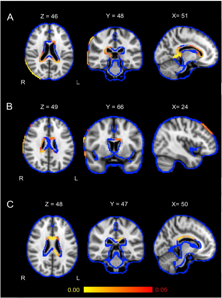
Methods: 72 participants (14 DLBs, 25 ADs, and 33 healthy controls (HCs)) underwent clinical assessment and 3 Tesla T1-weighted MRI at baseline and repeated at 12 months. We used FSL-SIENA to estimate PBVC for each subject. Voxelwise analyses and ANCOVA compared PBVC between DLB and AD, while correlational tests examined associations of PBVC with clinical measures.
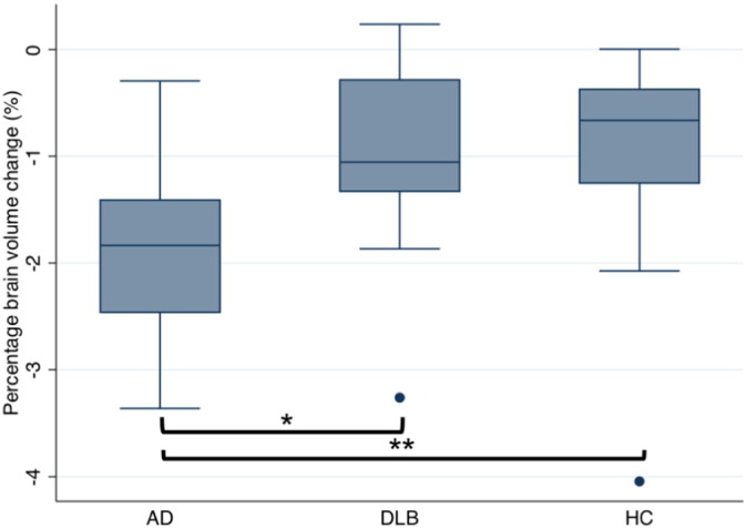
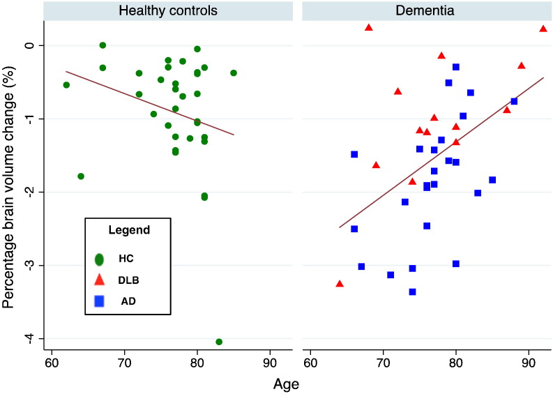
Results: AD had significantly greater atrophy over 1 year (1.8%) compared to DLB (1.0%; p = 0.01) and HC (0.9%; p < 0.01) in widespread regions of the brain including periventricular areas. PBVC was not significantly different between DLB and HC (p = 0.95). There were no differences in cognitive decline between DLB and AD. In the combined dementia group (AD and DLB), younger age was associated with higher atrophy rates (r = 0.49, p < 0.01).
Conclusions: AD showed a faster rate of global brain atrophy compared to DLB, which had similar rates of atrophy to HC. Among dementia subjects, younger age was associated with accelerated atrophy, reflecting more aggressive disease in younger people. PBVC could aid in differentiating between DLB and AD, however its utility as an outcome marker in DLB is limited.
Keywords: Alzheimer's disease; Atrophy; Dementia; Lewy bodies; Neuroimaging.
Figures
Similar articles
-
A comparison of medial and lateral temporal lobe atrophy in dementia with Lewy bodies and Alzheimer's disease: magnetic resonance imaging volumetric study.Dement Geriatr Cogn Disord. 2001 May-Jun;12(3):198-205. doi: 10.1159/000051258. Dement Geriatr Cogn Disord. 2001. PMID: 11244213
-
Temporal lobe atrophy on MRI in Parkinson disease with dementia: a comparison with Alzheimer disease and dementia with Lewy bodies.Neurology. 2005 Mar 8;64(5):861-5. doi: 10.1212/01.WNL.0000153070.82309.D4. Neurology. 2005. PMID: 15753423
-
Progressive cortical thinning and subcortical atrophy in dementia with Lewy bodies and Alzheimer's disease.Neurobiol Aging. 2015 Apr;36(4):1743-1750. doi: 10.1016/j.neurobiolaging.2014.12.038. Epub 2015 Jan 8. Neurobiol Aging. 2015. PMID: 25649023
-
[Differential diagnosis of dementia with lewy bodies].Brain Nerve. 2015 Apr;67(4):413-25. doi: 10.11477/mf.1416200157. Brain Nerve. 2015. PMID: 25846590 Review. Japanese.
-
How to diagnose dementia with Lewy bodies: state of the art.Mov Disord. 2005 Aug;20 Suppl 12:S11-20. doi: 10.1002/mds.20535. Mov Disord. 2005. PMID: 16092075 Review.
Cited by
-
Predicting Survival in Dementia With Lewy Bodies With Hippocampal Volumetry.Mov Disord. 2016 Jul;31(7):989-94. doi: 10.1002/mds.26666. Epub 2016 May 23. Mov Disord. 2016. PMID: 27214825 Free PMC article.
-
Cortical thinning in dementia with Lewy bodies and Parkinson disease dementia.Aust N Z J Psychiatry. 2020 Jun;54(6):633-643. doi: 10.1177/0004867419885165. Epub 2019 Nov 7. Aust N Z J Psychiatry. 2020. PMID: 31696728 Free PMC article.
-
Brain structure and cognitive ability in healthy aging: a review on longitudinal correlated change.Rev Neurosci. 2019 Dec 18;31(1):1-57. doi: 10.1515/revneuro-2018-0096. Rev Neurosci. 2019. PMID: 31194693 Free PMC article. Review.
-
Discrete pre-processing step effects in registration-based pipelines, a preliminary volumetric study on T1-weighted images.PLoS One. 2017 Oct 12;12(10):e0186071. doi: 10.1371/journal.pone.0186071. eCollection 2017. PLoS One. 2017. PMID: 29023597 Free PMC article.
-
Core outcome measures for interventions to prevent or slow the progress of dementia for people living with mild to moderate dementia: Systematic review and consensus recommendations.PLoS One. 2017 Jun 29;12(6):e0179521. doi: 10.1371/journal.pone.0179521. eCollection 2017. PLoS One. 2017. PMID: 28662127 Free PMC article.
References
-
- Ballmaier M., O'Brien J.T., Burton E.J., Thompson P.M., Rex D.E., Narr K.L., McKeith I.G., DeLuca H., Toga A.W. Comparing gray matter loss profiles between dementia with Lewy bodies and Alzheimer's disease using cortical pattern matching: diagnosis and gender effects. Neuroimage. 2004;23(1):325–335. 15325380 - PubMed
-
- Bartsch A.J., De Stefano N., Homola G., Smith S. Extending SIENA for a multi-subject statistical analysis of sample-specific cerebral edge shifts: substantiation of early brain regeneration through abstinence from alcoholism. Tenth Int. Conf. Funct. Mapp. Hum. Brain. 2004
-
- Burton E.J., Barber R., Mukaetova-Ladinska E.B., Robson J., Perry R.H., Jaros E., Kalaria R.N., O'Brien J.T. Medial temporal lobe atrophy on MRI differentiates Alzheimer's disease from dementia with Lewy bodies and vascular cognitive impairment: a prospective study with pathological verification of diagnosis. Brain. 2009;132(1):195–203. 19022858 - PubMed
Publication types
MeSH terms
LinkOut - more resources
Full Text Sources
Other Literature Sources
Medical
Molecular Biology Databases
