. 2015 Jan 27:7:482-92.
doi: 10.1016/j.nicl.2015.01.014. eCollection 2015.

White matter disruption at the prodromal stage of Alzheimer's disease: relationships with hippocampal atrophy and episodic memory performance

Affiliations

White matter disruption at the prodromal stage of Alzheimer's disease: relationships with hippocampal atrophy and episodic memory performance

Florence Rémy et al. Neuroimage Clin. .

Abstract

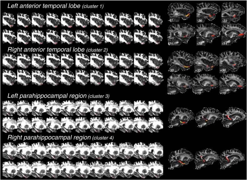
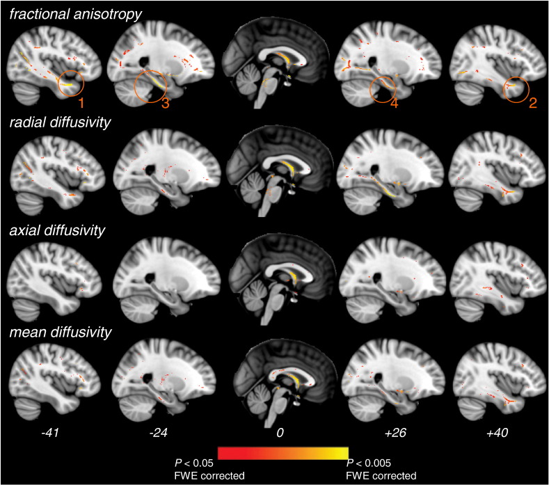
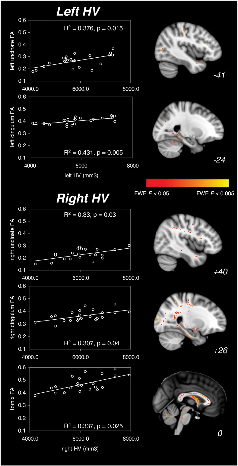
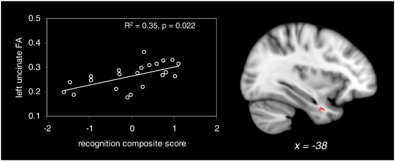
White matter tract alterations have been consistently described in Alzheimer's disease (AD). In particular, limbic fronto-temporal connections, which are critical to episodic memory function, may degenerate early in the course of the disease. However the relation between white matter tract degeneration, hippocampal atrophy and episodic memory impairment at the earliest stages of AD is still unclear. In this magnetic resonance imaging study, white matter integrity and hippocampal volumes were evaluated in patients with amnestic mild cognitive impairment due to AD (Albert et al., 2011) (n = 22) and healthy controls (n = 15). Performance in various episodic memory tasks was also evaluated in each participant. Relative to controls, patients showed a significant reduction of white matter fractional anisotropy (FA) and increase of radial diffusivity (RD) in the bilateral uncinate fasciculus, parahippocampal cingulum and fornix. Within the patient group, significant intra-hemispheric correlations were notably found between hippocampal grey matter volume and FA in the uncinate fasciculus, suggesting a relationship between atrophy and disconnection of the hippocampus. Moreover, episodic recognition scores were related with uncinate fasciculus FA across patients. These results indicate that fronto-hippocampal connectivity is reduced from the earliest pre-demential stages of AD. Disruption of fronto-hippocampal connections may occur progressively, in parallel with hippocampal atrophy, and may specifically contribute to early initial impairment in episodic memory.

Keywords: Alzheimer's disease; Cingulum; Diffusion tensor imaging; Episodic memory; Prodromal AD; Uncinate fasciculus.


Figures

Suppl. Fig. 1
Suppl. Fig. 1
Results of the back-projection of skeleton-space clusters evidenced in the group comparison analysis (Fig. 1) for the WM tracts of interest. Clusters in the left and right anterior temporal lobes (clusters 1 and 2 in Fig. 1, respectively) and in the left and right hippocampal formations (clusters 3 and 4 in Fig. 1, respectively) were back-projected onto individual FA images in standard space, for each subject. The left part of the figure shows the results of this back-projection for each patient (superimposed on individual FA images), confirming that skeleton clusters were derived from the correct tract-centre points in each of the patients. The right part of the figure shows the result of fibre tractography in a representative control subject, when taking each of the 4 clusters as a seed mask (see the Materials & methods section). The reconstructed tract is superimposed on the individual mean diffusion image in subject's native space. The approximate MNI × coordinate is indicated for each sagittal slice. The left and right uncinate fasciculi and the left and right parahippocampal cingula are clearly identified with fibre tractography.
Fig. 1
Fig. 1
Significant differences in DTI metrics between patients and controls. Regions of decreased fractional anisotropy and increased radial, axial and mean diffusivities in patients vs. controls are displayed. Medial temporal clusters showing significant FA decreases in patients are highlighted (orange circles). These clusters were further used as seed masks for tractography and as ROIs for correlation analyses within patients (see the Materials and methods section). All clusters shown are significant at p < 0.05 FWE-corrected for multiple comparisons using threshold-free cluster enhancement. DTI metrics and skeleton are superimposed on a standard T1 template. MNI x coordinates are indicated at the bottom of each slice.
Fig. 2
Fig. 2
Significant positive correlations between hippocampal volumes (HV) and FA measures in the patient group. The left part of the figure displays significant correlations between FA values in limbic tracts of interest and left and right hippocampal volumes. p-Values are Bonferroni-corrected for multiple comparisons. The right part of the figure shows whole-brain correlations between FA values and left HV (upper part) and right HV (bottom part) as a result of the TBSS analysis (p < 0.05 FWE-corrected). Significant clusters are superimposed on a standard T1 template. MNI x coordinates are indicated at the bottom of each sagittal plane.
Fig. 3
Fig. 3
Significant correlation between composite episodic recognition score and left uncinate fasciculus FA in the patient group (p < 0.05 Bonferroni-corrected for multiple comparisons). The right side of the figure shows the result of the whole-brain correlation between FA values and composite recognition scores. Significant clusters are superimposed on a standard T1 template. MNI x coordinate is indicated at the bottom of the sagittal slice.

Similar articles

Cited by

References

    1. Acosta-Cabronero J., Alley S., Williams G.B., Pengas G., Nestor P.J. Diffusion tensor metrics as biomarkers in Alzheimer's disease. PLOS One. 2012;7(11):e49072. - PMC - PubMed
    1. Acosta-Cabronero J., Williams G.B., Pengas G., Nestor P.J. Absolute diffusivities define the landscape of white matter degeneration in Alzheimer's disease. Brain. 2010;133(2):529–539. 19914928 - PubMed
    1. Albert M.S., DeKosky S.T., Dickson D., Dubois B., Feldman H.H., Fox N.C., Gamst A., Holtzman D.M., Jagust W.J., Petersen R.C., Snyder P.J., Carrillo M.C., Thies B., Phelps C.H. The diagnosis of mild cognitive impairment due to Alzheimer's disease: recommendations from the National Institute on Aging–Alzheimer's Association workgroups on diagnostic guidelines for Alzheimer's disease. Alzheimers Dement. 2011;7(3):270–279. - PMC - PubMed
    1. Allen G., Barnard H., McColl R., Hester A.L., Fields J.A., Weiner M.F., Ringe W.K., Lipton A.M., Brooker M., McDonald E., Rubin C.D., Cullum C.M. Reduced hippocampal functional connectivity in Alzheimer disease. Arch. Neurol. 2007;64(10):1482–1487. 17923631 - PubMed
    1. Bai F., Zhang Z., Watson D.R., Yu H., Shi Y., Yuan Y., Zang Y., Zhu C., Qian Y. Abnormal functional connectivity of hippocampus during episodic memory retrieval processing network in amnestic mild cognitive impairment. Biol. Psychiatry. 2009;65(11):951–958. 19028382 - PubMed

Publication types